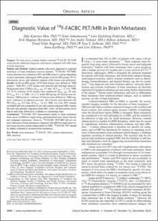
Diagnostic Value of 18F-FACBC PET/MRI in Brain Metastases
| dc.contributor.author | Øen, Silje Kjærnes | |
| dc.contributor.author | Johannessen, Knut | |
| dc.contributor.author | Pedersen, Lars Kjelsberg | |
| dc.contributor.author | Berntsen, Erik Magnus | |
| dc.contributor.author | Totland, Jon Andre | |
| dc.contributor.author | Johansen, Håkon | |
| dc.contributor.author | Bogsrud, Trond Velde | |
| dc.contributor.author | Solheim, Tora S | |
| dc.contributor.author | Karlberg, Anna Maria | |
| dc.contributor.author | Eikenes, Live | |
| dc.date.accessioned | 2023-01-16T12:19:14Z | |
| dc.date.available | 2023-01-16T12:19:14Z | |
| dc.date.created | 2022-12-02T09:51:37Z | |
| dc.date.issued | 2022 | |
| dc.identifier.citation | Clinical Nuclear Medicine. 2022, 47 (12), 1030-1039. | en_US |
| dc.identifier.issn | 0363-9762 | |
| dc.identifier.uri | https://hdl.handle.net/11250/3043713 | |
| dc.description.abstract | Purpose The study aims to evaluate whether combined 18F-FACBC PET/MRI could provide additional diagnostic information compared with MRI alone in brain metastases. Patients and Methods Eighteen patients with newly diagnosed or suspected recurrence of brain metastases received dynamic 18F-FACBC PET/MRI. Lesion detection was evaluated on PET and MRI scans in 2 groups depending on prior stereotactic radiosurgery (SRS group) or not (no-SRS group). SUVs, time-activity curves, and volumetric analyses of the lesions were performed. Results In the no-SRS group, 29/29 brain lesions were defined as “MRI positive.” With PET, 19/29 lesions were detected and had high tumor-to-background ratios (TBRs) (Dmax MR, ≥7 mm; SUVmax, 1.2–8.4; TBR, 3.9–25.9), whereas 10/29 lesions were undetected (Dmax MR, ≤8 mm; SUVmax, 0.3–1.2; TBR, 1.0–2.7). In the SRS group, 4/6 lesions were defined as “MRI positive,” whereas 2/6 lesions were defined as “MRI negative” indicative of radiation necrosis. All 6 lesions were detected with PET (Dmax MR, ≥15 mm; SUVmax, 1.4–4.2; TBR, 3.6–12.6). PET volumes correlated and were comparable in size with contrast-enhanced MRI volumes but were only partially congruent (mean DSC, 0.66). All time-activity curves had an early peak, followed by a plateau or a decreasing slope. Conclusions 18F-FACBC PET demonstrated uptake in brain metastases from cancer of different origins (lung, gastrointestinal tract, breast, thyroid, and malignant melanoma). However, 18F-FACBC PET/MRI did not improve detection of brain metastases compared with MRI but might detect tumor tissue beyond contrast enhancement on MRI. 18F-FACBC PET should be further evaluated in recurrent brain metastases. | en_US |
| dc.language.iso | eng | en_US |
| dc.publisher | Lippincott, Williams & Wilkins | en_US |
| dc.rights | Navngivelse-IkkeKommersiell-Ingen bearbeidelser 4.0 Internasjonal | |
| dc.rights.uri | http://creativecommons.org/licenses/by-nc-nd/4.0/deed.no | |
| dc.title | Diagnostic Value of 18F-FACBC PET/MRI in Brain Metastases | en_US |
| dc.title.alternative | Diagnostic Value of <sup>18</sup>F-FACBC PET/MRI in Brain Metastases | en_US |
| dc.type | Peer reviewed | en_US |
| dc.type | Journal article | en_US |
| dc.description.version | publishedVersion | en_US |
| dc.source.pagenumber | 1030-1039 | en_US |
| dc.source.volume | 47 | en_US |
| dc.source.journal | Clinical Nuclear Medicine | en_US |
| dc.source.issue | 12 | en_US |
| dc.identifier.doi | 10.1097/RLU.0000000000004435 | |
| dc.identifier.cristin | 2087588 | |
| cristin.ispublished | true | |
| cristin.fulltext | original | |
| cristin.qualitycode | 1 |
Tilhørende fil(er)
Denne innførselen finnes i følgende samling(er)

Med mindre annet er angitt, så er denne innførselen lisensiert som Navngivelse-IkkeKommersiell-Ingen bearbeidelser 4.0 Internasjonal