| dc.description.abstract | The modern western diet contains a high amount of omega-6 (n-6) polyunsaturated fatty acids (PUFAs) compared to omega-3 (n-3) PUFAs. Many studies have indicated that docosahexaenoic acid (DHA; 22:6 n-3) can inhibit cancer cell growth both in vivo and in vitro. Several mechanisms have been suggested to mediate the cytotoxic effect of n-3 PUFAs like alteration of prostaglandin synthesis, oxidative stress and lipid peroxidation, alterations in membrane composition, as well as transcription factors involved in signaling pathways like autophagy and the integrated stress response in cancer cells. Studies by our group have previously and recently shown that DHA induces ER stress and growth arrest in some human colorectal cancer cell lines. The aim of this thesis was to identify the molecular mechanisms involved in the anticancer properties of DHA in four human colorectal cancer cell lines, grown in the same culture medium, with special focus on basal autophagy level and the autophagy-regulating AKT-mTOR pathway.
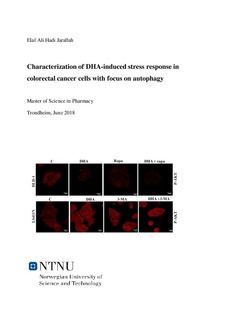
Four colorectal cancer cell lines, DLD-1, HCT-8, LS411N, and LS513 cells, responded differently to DHA (70 µM) treatment. All four cell lines were grown in the same growth medium. The effect of DHA on cell growth was estimated by cell counting, showing that DLD-1 and HCT-8 cells were sensitive to DHA (70 µM, 48 h) treatment with a growth inhibition of approximately 45 % and 25 % for DLD-1 and HCT-8 cells, respectively. LS411N and LS513 cells showed almost no growth inhibition given the same conditions. The basal level of autophagy and the level of protein MAP1LC3BII were found to be higher in the less DHA sensitive cell lines (LS411N and LS513) compared to DHA sensitive cell lines (DLD-1 and HCT-8). Gene expression analysis indicated an association between the level of DHA sensitivity and autophagy. DHA in combination with an autophagy inducer, rapamycin, decreased DHA sensitivity in DLD-1 and HCT-8 cells, while co-treatment with an autophagy inhibitor, 3-MA, increased the DHA sensitivity in LS411N and LS513 cells. Flow cytometry and the cyto-ID probe were used to assess the autophagic structures in the cells. The results indicated an increase in autophagic flux and the protein level of MAP1LC3BII in DLD-1 and HCT-8 cells after co- treatment with rapamycin and DHA, compared to rapamycin. In the LS411N and LS513 cells, the autophagic flux and level of MAP1LC3BII were found to decrease after co- treatment with 3-MA and DHA compared to 3-MA alone. The gene expression data indicated a possible connection between the AKT/mTOR pathway and the degree of DHA sensitivity in CRC cell lines. The p-AKT and p-mTOR levels decrease in DLD-1 and HCT-8 cells, indicating that the DHA-induced autophagy in DLD-1 and HCT-8 cells was possibly regulated by the AKT/mTOR pathway. Co-treatment with 3-MA and DHA was observed to increase the p-AKT and p-mTOR levels by confocal imaging, indicating a possible inhibition in autophagy level in LS411N and LS513 cells. Some of the results presented in figures 10 B, 15, and18 is recently published in FEBS Journal [1]. | nb_NO |