| dc.description.abstract | Blood and tissue motion can be detected using Doppler ultrasound. However, in a complex system such as the human body, the blood is not the only matter in motion. Doppler signals reflected by blood are often disturbed by stronger signals generated by the surrounding tissue, a phenomenon referred to as clutter. Clutter signals, being generally 40-100 dB stronger than the signals coming from blood, can usually be removed using conventional time filtering techniques. The problem arises when the surrounding tissue moves at velocities close to that of blood. In this case, conventional filters fail to remove the clutter components from the blood signal.
The aim of this thesis is to design and fabricate an in vitro phantom setup to evaluate ultrasound Doppler methods in coronary blood flow.
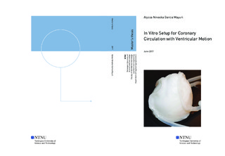
For this, we propose an experimental setup consisting of a polyvinyl alcohol (PVA) heart ventricle and coronary artery phantom including coronary blood flow circulation. The ventricle motion can be controlled with a pulsatile flow into the ventricle's cavity or by moving the ultrasound probe with a 3D positioning system.
PVA phantoms were fabricated with 12 hour and 24 hour freezing-thawing (F-T) cycle. The mechanical properties such as the speed of sound and shear wave velocity were measured. The results from the characterization of the PVA phantoms show that the sound speed was held almost constant for all F-T cycles, with a variation of only 0.5%. The shear wave velocity for the 12h sample increased 0.82 m/s/cycle, and 0.46 m/s/cycle for the sample with a 24h F-T cycle. The results showed a high experimental uncertainty which does not allow us to state a relation between the samples with a 12h and 24h F-T cycle.
Different scenarios for tissue motion controlled by a pulsatile flow and a robot were conducted to check the performance of a standard clutter filter using different pulse repetition frequencies (PRF): Including no tissue motion, a 40 bpm tissue motion, and a 70 bpm tissue motion. The results show that the chosen filter works perfectly for a PRF of 10000 Hz. For a PRF of 5000 Hz, the filter performs flawlessly, except for tissue motion at 70 bpm where some tissue signal remained unfiltered although with little effect in the Power Doppler (PD) image. For a PRF of 2000 Hz, tissue signal remained unfiltered using a 40 bpm and 70 bpm tissue motion.
Our results show that, as expected, the quality of blood flow visualization decreases with decreasing PRF.
For the robot controlled tissue motion, the clutter filter fails to remove the clutter components from the blood signal. The PD image of the filtered signal spectrum is all smeared out and the blood flow is not displayed.
We have managed to reproduce ventricular motion in a coronary circulation in vitro setup. More work must be done to check the repeatability and reproducibility of that motion, the setup opens the possibility to test advanced clutter filtering techniques in a controllable environment. | |