| dc.contributor.author | Knuth, Franziska Hanna | |
| dc.contributor.author | Grøndahl, Aurora Rosvoll | |
| dc.contributor.author | Winter, René | |
| dc.contributor.author | Torheim, Turid Katrine Gjerstad | |
| dc.contributor.author | Negård, Anne | |
| dc.contributor.author | Holmedal, Stein Harald | |
| dc.contributor.author | Bakke, Kine Mari | |
| dc.contributor.author | Meltzer, Sebastian | |
| dc.contributor.author | Futsæther, Cecilia Marie | |
| dc.contributor.author | Redalen, Kathrine | |
| dc.date.accessioned | 2023-01-26T10:10:29Z | |
| dc.date.available | 2023-01-26T10:10:29Z | |
| dc.date.created | 2022-05-12T14:14:14Z | |
| dc.date.issued | 2022 | |
| dc.identifier.citation | Physics and imaging in radiation oncology (PIRO). 2022, 22 77-84. | en_US |
| dc.identifier.issn | 2405-6316 | |
| dc.identifier.uri | https://hdl.handle.net/11250/3046534 | |
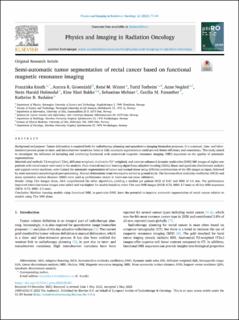
| dc.description.abstract | Background and purpose
Tumor delineation is required both for radiotherapy planning and quantitative imaging biomarker purposes. It is a manual, time- and labor-intensive process prone to inter- and intraobserver variations. Semi or fully automatic segmentation could provide better efficiency and consistency. This study aimed to investigate the influence of including and combining functional with anatomical magnetic resonance imaging (MRI) sequences on the quality of automatic segmentations.
Materials and methods
T2-weighted (T2w), diffusion weighted, multi-echo T2*-weighted, and contrast enhanced dynamic multi-echo (DME) MR images of eighty-one patients with rectal cancer were used in the analysis. Four classical machine learning algorithms; adaptive boosting (ADA), linear and quadratic discriminant analysis and support vector machines, were trained for automatic segmentation of tumor and normal tissue using different combinations of the MR images as input, followed by semi-automatic morphological post-processing. Manual delineations from two experts served as ground truth. The Sørensen-Dice similarity coefficient (DICE) and mean symmetric surface distance (MSD) were used as performance metric in leave-one-out cross validation.
Results
Using T2w images alone, ADA outperformed the other algorithms, yielding a median per patient DICE of 0.67 and MSD of 3.6 mm. The performance improved when functional images were added and was highest for models based on either T2w and DME images (DICE: 0.72, MSD: 2.7 mm) or all four MRI sequences (DICE: 0.72, MSD: 2.5 mm).
Conclusion
Machine learning models using functional MRI, in particular DME, have the potential to improve automatic segmentation of rectal cancer relative to models using T2w MRI alone. | en_US |
| dc.language.iso | eng | en_US |
| dc.publisher | Elsevier | en_US |
| dc.rights | Navngivelse 4.0 Internasjonal | * |
| dc.rights.uri | http://creativecommons.org/licenses/by/4.0/deed.no | * |
| dc.title | Semi-automatic tumor segmentation of rectal cancer based on functional magnetic resonance imaging | en_US |
| dc.title.alternative | Semi-automatic tumor segmentation of rectal cancer based on functional magnetic resonance imaging | en_US |
| dc.type | Peer reviewed | en_US |
| dc.type | Journal article | en_US |
| dc.description.version | publishedVersion | en_US |
| dc.source.pagenumber | 77-84 | en_US |
| dc.source.volume | 22 | en_US |
| dc.source.journal | Physics and imaging in radiation oncology (PIRO) | en_US |
| dc.identifier.doi | 10.1016/j.phro.2022.05.001 | |
| dc.identifier.cristin | 2023977 | |
| dc.relation.project | Samarbeidsorganet mellom Helse Midt-Norge og NTNU: 30513 | en_US |
| dc.relation.project | Kreftforeningen: 198116-2018 | en_US |
| dc.relation.project | Helse Sør-Øst RHF: 2013002 | en_US |
| dc.relation.project | Helse Sør-Øst RHF: 2015048 | en_US |
| dc.relation.project | Helse Sør-Øst RHF: 2016050 | en_US |
| cristin.ispublished | true | |
| cristin.fulltext | original | |
| cristin.qualitycode | 1 | |

