| dc.contributor.author | Randeberg, Lise Lyngsnes | |
| dc.contributor.author | Hegstad, Janne-Lise A | |
| dc.contributor.author | Paluchowski, Lukasz A. | |
| dc.contributor.author | Milanic, Matija | |
| dc.contributor.author | Pukstad, Brita | |
| dc.date.accessioned | 2015-01-18T12:07:12Z | |
| dc.date.accessioned | 2015-02-10T14:49:53Z | |
| dc.date.available | 2015-01-18T12:07:12Z | |
| dc.date.available | 2015-02-10T14:49:53Z | |
| dc.date.issued | 2014 | |
| dc.identifier.citation | Proceedings of SPIE, the International Society for Optical Engineering 2014, 8926 | nb_NO |
| dc.identifier.issn | 0277-786X | |
| dc.identifier.uri | http://hdl.handle.net/11250/275803 | |
| dc.description | This is the author’s final, accepted and refereed manuscript to the article. | nb_NO |
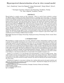
| dc.description.abstract | Wound healing is a complex process not fully understood. There is a need of better methods to evaluate the different stages of healing, and optical characterization is a promising tool in this respect. In this study hyperspectral imaging was employed to characterize an in vitro wound model. The wound model was established by first cutting circular patches of human abdominal skin using an 8mm punch biopsy tool, and then creating dermal wounds in the center of the skin patches using a 5mm tool. The wounds were incubated in medium with 10% serum and antibiotics. Hyperspectral images were collected every three days using a push broom hyper spectral camera (Hyspex VNIR1600). The camera had a spectral resolution of 3.7 nm and was fitted with a close up lens giving a FOV of 2.5 cm and a spatial resolution of 29 micrometer. Samples for histology were collected throughout the measurement period, which was 21 days in total. Data were processed in ENVI and Matlab. A successful classification based on hyperspectral imaging of the implemented model is presented. It was not possible to see the healing zone in the in vitro model with the naked eye without dying. The hyperspectral results showed that newly formed epithelium could be imaged without any additional contrast agents or dyes. It was also possible to detect non-viable tissue. In vitro wound models and hyperspectral imaging can thus be employed to gain further insight in the complicated process of healing in different kinds of wounds. © (2014) COPYRIGHT Society of Photo-Optical Instrumentation Engineers (SPIE). Downloading of the abstract is permitted for personal use only. | nb_NO |
| dc.language.iso | eng | nb_NO |
| dc.publisher | SPIE | nb_NO |
| dc.title | Hyperspectral characterization of an in vitro wound model | nb_NO |
| dc.type | Journal article | nb_NO |
| dc.type | Peer reviewed | en_GB |
| dc.date.updated | 2015-01-18T12:07:12Z | |
| dc.source.volume | 8926 | nb_NO |
| dc.source.journal | Proceedings of SPIE, the International Society for Optical Engineering | nb_NO |
| dc.identifier.doi | 10.1117/12.2039976 | |
| dc.identifier.cristin | 1200221 | |
| dc.description.localcode | Copyright 2014. Society of Photo Optical Instrumentation Engineers. One print or electronic copy may be made for personal use only. Systematic reproduction and distribution, duplication of any material in this paper for a fee or for commercial purposes, or modification of the content of the paper are prohibited. | nb_NO |