Hippocampal-medial entorhinal circuit is differently organized along the dorsoventral axis in rodents
| dc.contributor.author | Ohara, Shinya | |
| dc.contributor.author | Rannap, Märt | |
| dc.contributor.author | Tsutsui, Ken-Ichiro | |
| dc.contributor.author | Draguhn, Andreas | |
| dc.contributor.author | Egorov, Alexei V. | |
| dc.contributor.author | Witter, Menno Peter | |
| dc.date.accessioned | 2023-10-26T13:10:57Z | |
| dc.date.available | 2023-10-26T13:10:57Z | |
| dc.date.created | 2023-03-06T09:55:26Z | |
| dc.date.issued | 2023 | |
| dc.identifier.citation | Cell reports. 2023, 42 (1), . | en_US |
| dc.identifier.issn | 2211-1247 | |
| dc.identifier.uri | https://hdl.handle.net/11250/3098971 | |
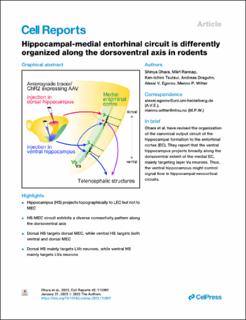
| dc.description.abstract | The general understanding of hippocampal circuits is that the hippocampus and the entorhinal cortex (EC) are topographically connected through parallel identical circuits along the dorsoventral axis. Our anterograde tracing and in vitro electrophysiology data, however, show a markedly different dorsoventral organization of the hippocampal projection to the medial EC (MEC). While dorsal hippocampal projections are confined to the dorsal MEC, ventral hippocampal projections innervate both dorsal and ventral MEC. Further, whereas the dorsal hippocampus preferentially targets layer Vb (LVb) neurons, the ventral hippocampus mainly targets cells in layer Va (LVa). This connectivity scheme differs from hippocampal projections to the lateral EC, which are topographically organized along the dorsoventral axis. As LVa neurons project to telencephalic structures, our findings indicate that the ventral hippocampus regulates LVa-mediated entorhinal-neocortical output from both dorsal and ventral MEC. Overall, the marked dorsoventral differences in hippocampal-entorhinal connectivity impose important constraints on signal flow in hippocampal-neocortical circuits. | en_US |
| dc.language.iso | eng | en_US |
| dc.publisher | Elsevier | en_US |
| dc.rights | Navngivelse 4.0 Internasjonal | * |
| dc.rights.uri | http://creativecommons.org/licenses/by/4.0/deed.no | * |
| dc.title | Hippocampal-medial entorhinal circuit is differently organized along the dorsoventral axis in rodents | en_US |
| dc.title.alternative | Hippocampal-medial entorhinal circuit is differently organized along the dorsoventral axis in rodents | en_US |
| dc.type | Peer reviewed | en_US |
| dc.type | Journal article | en_US |
| dc.description.version | publishedVersion | en_US |
| dc.source.pagenumber | 0 | en_US |
| dc.source.volume | 42 | en_US |
| dc.source.journal | Cell reports | en_US |
| dc.source.issue | 1 | en_US |
| dc.identifier.doi | 10.1016/j.celrep.2023.112001 | |
| dc.identifier.cristin | 2131410 | |
| cristin.ispublished | true | |
| cristin.fulltext | original | |
| cristin.qualitycode | 2 |
Files in this item
This item appears in the following Collection(s)

Except where otherwise noted, this item's license is described as Navngivelse 4.0 Internasjonal