Biomechanics of mitral valve leaflets: Second harmonic generation microscopy, biaxial mechanical tests and tissue modeling
| dc.contributor.author | Sadeghinia, MohammadJavad | |
| dc.contributor.author | Skallerud, Bjørn Helge | |
| dc.contributor.author | Holzapfel, Gerhard | |
| dc.contributor.author | Prot, Victorien Emile | |
| dc.date.accessioned | 2023-02-27T12:03:33Z | |
| dc.date.available | 2023-02-27T12:03:33Z | |
| dc.date.created | 2022-08-30T09:04:15Z | |
| dc.date.issued | 2022 | |
| dc.identifier.citation | Acta Biomaterialia. 2022, 141 244-254. | en_US |
| dc.identifier.issn | 1742-7061 | |
| dc.identifier.uri | https://hdl.handle.net/11250/3054208 | |
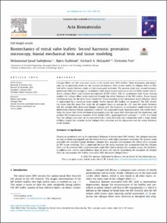
| dc.description.abstract | Collagen fibers are the main load carrier in the mitral valve (MV) leaflets. Their orientation and dispersion are an important factor for the mechanical behavior. Most recent studies of collagen fibers in MVs lack either entire thickness study or high transmural resolution. The present study uses second harmonic generation (SHG) microscopy in combination with planar biaxial mechanical tests to better model and examine collagen fibers and mechanical properties of MV leaflets. SHG in combination with tissue clearing enables the collagen fibers to be examined through the entire thickness of the MV leaflets. Planar biaxial mechanical tests, on the other hand, enable the characterization of the mechanical tissue behavior, which is represented by a structural tissue model. Twelve porcine MV leaflets are examined. The SHG recording shows that the mean fiber angle for all samples varies on average by ±12° over the entire thickness and the collagen fiber dispersion changes strongly over the thickness. A constitutive model based on the generalized structure tensor approach is used for the associated tissue characterization. The model represents the tissue with three mechanical parameters plus the mean fiber direction and the dispersion, and predicts the biomechanical response of the leaflets with a good agreement (average ). It is found that the collagen structure can be represented by a mean direction and a dispersion with a single family of fibers despite the variation in the collagen fiber direction and the dispersion over the entire thickness of MV leaflets. | en_US |
| dc.language.iso | eng | en_US |
| dc.publisher | Elsevier | en_US |
| dc.rights | Navngivelse 4.0 Internasjonal | * |
| dc.rights.uri | http://creativecommons.org/licenses/by/4.0/deed.no | * |
| dc.title | Biomechanics of mitral valve leaflets: Second harmonic generation microscopy, biaxial mechanical tests and tissue modeling | en_US |
| dc.title.alternative | Biomechanics of mitral valve leaflets: Second harmonic generation microscopy, biaxial mechanical tests and tissue modeling | en_US |
| dc.type | Peer reviewed | en_US |
| dc.type | Journal article | en_US |
| dc.description.version | publishedVersion | en_US |
| dc.source.pagenumber | 244-254 | en_US |
| dc.source.volume | 141 | en_US |
| dc.source.journal | Acta Biomaterialia | en_US |
| dc.identifier.doi | 10.1016/j.actbio.2022.01.003 | |
| dc.identifier.cristin | 2046986 | |
| cristin.ispublished | true | |
| cristin.fulltext | original | |
| cristin.qualitycode | 1 |
Tilhørende fil(er)
Denne innførselen finnes i følgende samling(er)

Med mindre annet er angitt, så er denne innførselen lisensiert som Navngivelse 4.0 Internasjonal