| dc.contributor.author | Halset, Jørgen Holli | |
| dc.contributor.author | Hanssen, Simon Wøhlert | |
| dc.contributor.author | Espinosa, Aurora | |
| dc.contributor.author | Klepstad, Pål | |
| dc.date.accessioned | 2015-09-01T08:08:39Z | |
| dc.date.accessioned | 2015-09-03T08:11:14Z | |
| dc.date.available | 2015-09-01T08:08:39Z | |
| dc.date.available | 2015-09-03T08:11:14Z | |
| dc.date.issued | 2015 | |
| dc.identifier.citation | BMC Anesthesiology 2015, 15(28) | nb_NO |
| dc.identifier.issn | 1471-2253 | |
| dc.identifier.uri | http://hdl.handle.net/11250/298522 | |
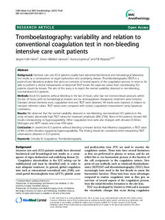
| dc.description.abstract | Background: Intensive care unit (ICU) patients usually have abnormal biochemical and hematological laboratory
test results as a consequence of organ dysfunction and underlying disease. Thromboelastography (TEG®) is a
point-of-care laboratory analysis that gives an overview of several aspects of the coagulation process. In order to be
able to perform a clinical interpretation of abnormal TEG® results the expected values from non-bleeding ICU
patients should be known. The aim of this study is to report the normal variability observed in non-bleeding,
non-transfused ICU patients.
Methods: Adult ICU patients without bleeding in the last 24 hours, who had not received blood products within
the last 24 hours, with no hematological diseases and no anticoagulation therapeutic treatment were included.
Standard clinical chemistry tests, coagulation tests and TEG® were obtained. All results were reported in relation to
standard reference values. TEG® values were compared with routine coagulation measurement using Spearman
correlations.
Results: We observed that the normal variability observed in non-bleeding, non-transfused ICU patients in this
study included abnormally high TEG® values for maximum amplitude (MA) (73%). None of the patients showed MA
results corresponding to hypocoagulability. Other coagulation tests were also changed with elevated D-Dimer,
fibrinogen and APTT values, and a low ATIII value.
Conclusion: In unselected ICU patients without bleeding or known factors that influence coagulation, a TEG® value
of MA is often elevated suggesting hypercoagulability. This finding should be considered when interpreting TEG®
observations obtained in ICU patients.
Keywords: Critically ill, Coagulation, Tromboelastography | nb_NO |
| dc.language.iso | eng | nb_NO |
| dc.publisher | BioMed Central | nb_NO |
| dc.title | Tromboelastography: variability and relation to conventional coagulation test in non-bleeding intensive care unit patients | nb_NO |
| dc.type | Journal article | nb_NO |
| dc.type | Peer reviewed | en_GB |
| dc.date.updated | 2015-09-01T08:08:39Z | |
| dc.source.volume | 15 | nb_NO |
| dc.source.journal | BMC Anesthesiology | nb_NO |
| dc.source.issue | 28 | nb_NO |
| dc.identifier.doi | 10.1186/s12871-015-0011-2 | |
| dc.identifier.cristin | 1261127 | |
| dc.description.localcode | © 2015 Holli Halset et al.; licensee BioMed Central. This is an Open Access article distributed under the terms of the Creative Commons Attribution License (http://creativecommons.org/licenses/by/4.0), which permits unrestricted use, distribution, and reproduction in any medium, provided the original work is properly credited. The Creative Commons Public Domain Dedication waiver (http://creativecommons.org/publicdomain/zero/1.0/) applies to the data made available in this article, unless otherwise stated. | nb_NO |
