Fetal molding examined with transperineal ultrasound and associations with position and delivery mode
| dc.contributor.author | Iversen, Johanne Kolvik | |
| dc.contributor.author | Kahrs, Birgitte Heiberg | |
| dc.contributor.author | Torkildsen, Erik A. | |
| dc.contributor.author | Eggebø, Torbjørn Moe | |
| dc.date.accessioned | 2022-02-28T14:27:28Z | |
| dc.date.available | 2022-02-28T14:27:28Z | |
| dc.date.created | 2020-12-11T17:01:33Z | |
| dc.date.issued | 2020 | |
| dc.identifier.citation | American Journal of Obstetrics and Gynecology. 2020, . | en_US |
| dc.identifier.issn | 0002-9378 | |
| dc.identifier.uri | https://hdl.handle.net/11250/2981791 | |
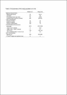
| dc.description.abstract | Background To accommodate passage through the birth canal, the fetal skull is compressed and reshaped, a phenomenon known as molding. The fetal skull bones are separated by membranous sutures that facilitate compression and overlap, resulting in a reduced diameter. This increases the probability of a successful vaginal delivery. Fetal position, presentation, station, and attitude can be examined with ultrasound, but fetal head molding has not been previously studied with ultrasound. Objective This study aimed to describe ultrasound-assessed fetal head molding in a population of nulliparous women with slow progress in the second stage of labor and to study associations with fetal position and delivery mode. Study Design This was a secondary analysis of a population comprising 150 nulliparous women with a single fetus in cephalic presentation, with slow progress in the active second stage with pushing. Women were eligible for the study when an operative intervention was considered by the clinician. Molding was examined in stored transperineal two-dimensional and three-dimensional acquisitions and differentiated into occipitoparietal molding along the lambdoidal sutures, frontoparietal molding along the coronal sutures, and parietoparietal molding at the sagittal suture (molding in the midline). Molding could not be classified if positions were unknown, and these cases were excluded. We measured the distance from the molding to the head midline, molding step, and overlap of skull bones and looked for associations with fetal position and delivery mode. The responsible clinicians were blinded to the ultrasound findings. Results Six cases with unknown position were excluded, leaving 144 women in the study population. Fetal position was anterior in 117 cases, transverse in 12 cases, and posterior in 15 cases. Molding was observed in 79 of 144 (55%) fetuses. Molding was seen significantly more often in occiput anterior positions than in non–occiput anterior positions (69 of 117 [59%] vs 10 of 27 [37%]; P=.04). In occiput anterior positions, the molding was seen as occipitoparietal molding in 68 of 69 cases and as parietoparietal molding in 1 case with deflexed attitude. Molding was seen in 19 of 38 (50%) of occiput anterior positions ending with spontaneous delivery, 42 of 71(59%) ending with vacuum extraction, and in 7 of 8 (88%) with failed vacuum extraction (P=.13). In 4 fetuses with occiput posterior positions, parietoparietal molding was diagnosed, and successful vacuum extraction occurred in 3 cases and failed extraction in 1. Frontoparietal molding was seen in 2 transverse positions and 4 posterior positions. One delivered spontaneously; vacuum extraction failed in 3 cases and was successful in 2. Only 1 of 11 fetuses with either parietoparietal or frontoparietal molding was delivered spontaneously. Conclusion The different types of molding can be classified with ultrasound. Occipitoparietal molding was commonly seen in occiput anterior positions and not significantly associated with delivery mode. Frontoparietal and parietoparietal moldings were less frequent than reported in old studies and should be studied in larger populations with mixed ethnicities. | en_US |
| dc.language.iso | eng | en_US |
| dc.publisher | Elsevier | en_US |
| dc.rights.uri | https://creativecommons.org/licenses/by-nc-nd/4.0/ | |
| dc.title | Fetal molding examined with transperineal ultrasound and associations with position and delivery mode | en_US |
| dc.type | Journal article | en_US |
| dc.type | Peer reviewed | en_US |
| dc.description.version | acceptedVersion | en_US |
| dc.rights.holder | © 2020. This manuscript version is made available under the CC-BY-NC-ND 4.0 license https://creativecommons.org/licenses/by-nc-nd/4.0/ | en_US |
| dc.source.pagenumber | 0 | en_US |
| dc.source.journal | American Journal of Obstetrics and Gynecology | en_US |
| dc.identifier.doi | 10.1016/j.ajog.2020.06.042 | |
| dc.identifier.cristin | 1858890 | |
| dc.description.localcode | Not available due to copyright restrictions | en_US |
| cristin.ispublished | true | |
| cristin.fulltext | postprint | |
| cristin.fulltext | postprint | |
| cristin.qualitycode | 2 |
Tilhørende fil(er)
Denne innførselen finnes i følgende samling(er)

Med mindre annet er angitt, så er denne innførselen lisensiert som https://creativecommons.org/licenses/by-nc-nd/4.0/