| dc.contributor.author | Arnli, Magnus B. | |
| dc.contributor.author | Backer-Grøndahl, Thomas | |
| dc.contributor.author | Ytterhus, Borgny | |
| dc.contributor.author | Granli, Unn S. | |
| dc.contributor.author | Lydersen, Stian | |
| dc.contributor.author | Gulati, Sasha | |
| dc.contributor.author | Torp, Sverre Helge | |
| dc.date.accessioned | 2017-04-06T07:50:23Z | |
| dc.date.available | 2017-04-06T07:50:23Z | |
| dc.date.created | 2017-03-29T22:40:23Z | |
| dc.date.issued | 2017 | |
| dc.identifier.issn | 2167-8359 | |
| dc.identifier.uri | http://hdl.handle.net/11250/2436934 | |
| dc.description.abstract | Background
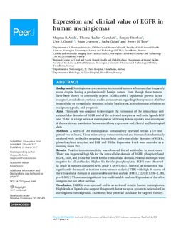
Meningiomas are common intracranial tumors in humans that frequently recur despite having a predominantly benign nature. Even though these tumors have been shown to commonly express EGFR/c-erbB1 (epidermal growth factor receptor), results from previous studies are uncertain regarding the expression of either intracellular or extracellular domains, cellular localization, activation state, relations to malignancy grade, and prognosis.
Aims
This study was designed to investigate the expression of the intracellular and extracellular domains of EGFR and of the activated receptor as well as its ligands EGF and TGFα in a large series of meningiomas with long follow-up data, and investigate if there exists an association between antibody expression and clinical and histological data.
Methods
A series of 186 meningiomas consecutively operated within a 10-year period was included. Tissue microarrays were constructed and immunohistochemically analyzed with antibodies targeting intracellular and extracellular domains of EGFR, phosphorylated receptor, and EGF and TGFα. Expression levels were recorded as a staining index (SI).
Results
Positive immunoreactivity was observed for all antibodies in most cases. There was in general high SIs for the intracellular domain of EGFR, phosphorylated EGFR, EGF, and TGFα but lower for the extracellular domain. Normal meninges were negative for all antibodies. Higher SIs for the phosphorylated EGFR were observed in grade II tumors compared with grade I (p = 0.018). Survival or recurrence was significantly decreased in the time to recurrence analysis (TTR) with high SI-scores of the extracellular domain in a univariable survival analysis (HR 1.152, CI (1.036–1.280, p = 0.009)). This was not significant in a multivariable analysis. Expression of the other antigens did not affect survival.
Conclusion
EGFR is overexpressed and in an activated state in human meningiomas. High levels of ligands also support this growth factor receptor system to be involved in meningioma tumorigenesis. EGFR may be a potential candidate for targeted therapy. | nb_NO |
| dc.language.iso | eng | nb_NO |
| dc.publisher | PeerJ | nb_NO |
| dc.rights | Navngivelse 4.0 Internasjonal | * |
| dc.rights.uri | http://creativecommons.org/licenses/by/4.0/deed.no | * |
| dc.title | Expression and clinical value of EGFR in human meningiomas | nb_NO |
| dc.type | Journal article | nb_NO |
| dc.type | Peer reviewed | nb_NO |
| dc.source.volume | 5 | nb_NO |
| dc.source.journal | PeerJ | nb_NO |
| dc.identifier.doi | https://doi.org/10.7717/peerj.3140 | |
| dc.identifier.cristin | 1462226 | |
| dc.description.localcode | Copyright © 2017 Arnli et al. This is an open access article distributed under the terms of the Creative Commons Attribution License, which permits unrestricted use, distribution, reproduction and adaptation in any medium and for any purpose provided that it is properly attributed. For attribution, the original author(s), title, publication source (PeerJ) and either DOI or URL of the article must be cited. | nb_NO |
| cristin.unitcode | 194,65,10,0 | |
| cristin.unitcode | 194,65,0,0 | |
| cristin.unitcode | 194,65,35,0 | |
| cristin.unitname | Institutt for laboratoriemedisin, barne- og kvinnesykdommer | |
| cristin.unitname | Det medisinske fakultet | |
| cristin.unitname | Regionalt kunnskapssenter for barn og unge - Psykisk helse og barnevern | |
| cristin.ispublished | true | |
| cristin.fulltext | original | |
| cristin.qualitycode | 1 | |

