PHH3 in breast cancer - Using immunohistochemistry to visualize mitoses
Master thesis
Permanent lenke
http://hdl.handle.net/11250/2417719Utgivelsesdato
2016Metadata
Vis full innførselSamlinger
Sammendrag
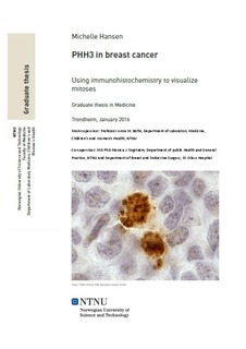
Mitotic activity is an independent prognostic indicator in breast cancer and is one of the three morphological characteristics assessed when determining histopathological grade. However, mitotic counts carried out on routinely stained sections are time-consuming and may be subject to inter- and intra-observer variation. In this study we compared mitotic counts performed on routine, haematoxylin-erythrosine-saffron (HES) stained sections with mitotic counts carried out on sections stained by immunohistochemistry for the mitotic marker Anti-Phospho-Histone H3 (Ser10)(PHH3), a marker of cells in late G2 to M phase. The study population comprised tissue samples from 250 cases of symptomatic breast cancer assembled in tissue microarrays (TMA). All cases were stained with HES and PHH3 and mitoses were counted. Mitotic counts carried out on HES-stained whole sections from the same cases were also available. We found that mitotic counts in PHH3-stained slides were consistently higher compared to HES-stained slides. However, the results showed good correlation between the methods (0.72 by Pearson correlation test). PHH3 counts correlated well with histopathological grade and molecular subtypes. No clear association with breast cancer specific survival was observed. The number of cases is relatively low and TMA is an additional limitation. However, the results of this study indicate that further studies of PHH3 as an alternative method of visualizing mitoses are warranted.