| dc.contributor.author | Blogg, Lesley S | |
| dc.contributor.author | Gennser, Mikael | |
| dc.contributor.author | Møllerløkken, Andreas | |
| dc.contributor.author | Brubakk, Alf O | |
| dc.date.accessioned | 2015-02-06T13:21:58Z | |
| dc.date.accessioned | 2016-09-08T13:18:58Z | |
| dc.date.available | 2015-02-06T13:21:58Z | |
| dc.date.available | 2016-09-08T13:18:58Z | |
| dc.date.issued | 2014 | |
| dc.identifier.citation | Diving and Hyperbaric Medicine 2014, 44(1):35-44 | nb_NO |
| dc.identifier.issn | 1833-3516 | |
| dc.identifier.uri | http://hdl.handle.net/11250/2405515 | |
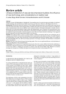
| dc.description.abstract | INTRODUCTION:
Diving often causes the formation of 'silent' bubbles upon decompression. If the bubble load is high, then the risk of decompression sickness (DCS) and the number of bubbles that could cross to the arterial circulation via a pulmonary shunt or patent foramen ovale increase. Bubbles can be monitored aurally, with Doppler ultrasound, or visually, with two dimensional (2D) ultrasound imaging. Doppler grades and imaging grades can be compared with good agreement. Early 2D imaging units did not provide such comprehensive observations as Doppler, but advances in technology have allowed development of improved, portable, relatively inexpensive units. Most now employ harmonic technology; it was suggested that this could allow previously undetectable bubbles to be observed.
METHODS:
This paper provides a review of current methods of bubble measurement and how new technology may be changing our perceptions of the potential relationship of these measurements to decompression illness. Secondly, 69 paired ultrasound images were made using conventional 2D ultrasound imaging and harmonic imaging. Images were graded on the Eftedal-Brubakk (EB) scale and the percentage agreement of the images calculated. The distribution of mismatched grades was analysed.
RESULTS:
Fifty-four of the 69 paired images had matching grades. There was no significant difference in the distribution of high or low EB grades for the mismatched pairs.
CONCLUSIONS:
Given the good level of agreement between pairs observed, it seems unlikely that harmonic technology is responsible for any perceived increase in observed bubble loads, but it is probable that our increasing use of 2D ultrasound to assess dive profiles is changing our perception of 'normal' venous and arterial bubble loads. Methods to accurately investigate the load and size of bubbles developed will be helpful in the future in determining DCS risk. | nb_NO |
| dc.language.iso | eng | nb_NO |
| dc.publisher | South Pacific Medicine Society and the European Underwater and Baromedical Society. | nb_NO |
| dc.subject | Doppler; arterial gas embolism; bubbles; decompression sickness; diving research; review article; venous gas embolism | nb_NO |
| dc.title | Ultrasound detection of vascular decompression bubbles: The influence of new technology and considerations on bubble load | nb_NO |
| dc.type | Journal article | nb_NO |
| dc.type | Peer reviewed | nb_NO |
| dc.date.updated | 2015-02-06T13:21:58Z | |
| dc.source.pagenumber | 35-44 | nb_NO |
| dc.source.volume | 44 | nb_NO |
| dc.source.journal | Diving and Hyperbaric Medicine | nb_NO |
| dc.source.issue | 1 | nb_NO |
| dc.identifier.cristin | 1128146 | |
| dc.description.localcode | Author postprint | nb_NO |
