MRI Reveals the in Vivo Cellular and Vascular Response to BEZ235 in Ovarian Cancer Xenografts with Different PI3-Kinase Pathway Activity
| dc.contributor.author | Cebulla, Jana | |
| dc.contributor.author | Huuse-Røneid, Else Marie | |
| dc.contributor.author | Pettersen, Kristine | |
| dc.contributor.author | van der Veen, Anna | |
| dc.contributor.author | Kim, Eugene | |
| dc.contributor.author | Andersen, Sonja | |
| dc.contributor.author | Prestvik, Wenche S | |
| dc.contributor.author | Bofin, Anna M. | |
| dc.contributor.author | Pathak, Arvind P | |
| dc.contributor.author | Bjørkøy, Geir | |
| dc.contributor.author | Bathen, Tone Frost | |
| dc.contributor.author | Moestue, Siver Andreas | |
| dc.date.accessioned | 2016-08-31T08:15:23Z | |
| dc.date.accessioned | 2016-09-08T11:17:50Z | |
| dc.date.available | 2016-08-31T08:15:23Z | |
| dc.date.available | 2016-09-08T11:17:50Z | |
| dc.date.issued | 2014-12-23 | |
| dc.identifier.citation | British Journal of Cancer 2015, 112(3):504-513 | nb_NO |
| dc.identifier.issn | 1532-1827 | |
| dc.identifier.uri | http://hdl.handle.net/11250/2405396 | |
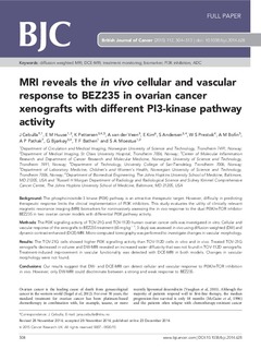
| dc.description.abstract | Background: The phosphoinositide-3 kinase (PI3K) pathway is an attractive therapeutic target. However, difficulty in predicting therapeutic response limits the clinical implementation of PI3K inhibitors. This study evaluates the utility of clinically relevant magnetic resonance imaging (MRI) biomarkers for noninvasively assessing the in vivo response to the dual PI3K/mTOR inhibitor BEZ235 in two ovarian cancer models with differential PI3K pathway activity. Methods: The PI3K signalling activity of TOV-21G and TOV-112D human ovarian cancer cells was investigated in vitro. Cellular and vascular response of the xenografts to BEZ235 treatment (65 mg kg−1, 3 days) was assessed in vivo using diffusion-weighted (DW) and dynamic contrast-enhanced (DCE)-MRI. Micro-computed tomography was performed to investigate changes in vascular morphology. Results: The TOV-21G cells showed higher PI3K signalling activity than TOV-112D cells in vitro and in vivo. Treated TOV-21G xenografts decreased in volume and DW-MRI revealed an increased water diffusivity that was not found in TOV-112D xenografts. Treatment-induced improvement in vascular functionality was detected with DCE-MRI in both models. Changes in vascular morphology were not found. Conclusions: Our results suggest that DW- and DCE-MRI can detect cellular and vascular response to PI3K/mTOR inhibition in vivo. However, only DW-MRI could discriminate between a strong and weak response to BEZ235. | nb_NO |
| dc.language.iso | eng | nb_NO |
| dc.publisher | Cancer Research UK | nb_NO |
| dc.rights | Navngivelse-Ikkekommersiell-DelPåSammeVilkår 3.0 Norge | * |
| dc.rights.uri | http://creativecommons.org/licenses/by-nc-sa/3.0/no/ | * |
| dc.title | MRI Reveals the in Vivo Cellular and Vascular Response to BEZ235 in Ovarian Cancer Xenografts with Different PI3-Kinase Pathway Activity | nb_NO |
| dc.type | Journal article | nb_NO |
| dc.type | Peer reviewed | nb_NO |
| dc.date.updated | 2016-08-31T08:15:23Z | |
| dc.source.pagenumber | 504-513 | nb_NO |
| dc.source.volume | 112 | nb_NO |
| dc.source.journal | British Journal of Cancer | nb_NO |
| dc.source.issue | 3 | nb_NO |
| dc.identifier.doi | 10.1038/bjc.2014.628 | |
| dc.identifier.cristin | 1183726 | |
| dc.relation.project | Norges forskningsråd: 223255 | nb_NO |
| dc.description.localcode | From twelve months after its original publication, this work is licensed under the Creative Commons Attribution-NonCommercial-Share Alike 3.0 Unported License. | nb_NO |
Files in this item
This item appears in the following Collection(s)

Except where otherwise noted, this item's license is described as Navngivelse-Ikkekommersiell-DelPåSammeVilkår 3.0 Norge