| dc.contributor.author | Jannig, Paulo R. | |
| dc.contributor.author | Moreira, Jose Bianco Nascimento | |
| dc.contributor.author | Bechara, Luiz R.G. | |
| dc.contributor.author | Bozi, Luiz H.M. | |
| dc.contributor.author | Bacurau, Aline V. | |
| dc.contributor.author | Monteiro, Alex W.A. | |
| dc.contributor.author | Dourado, Paulo M. | |
| dc.contributor.author | Wisløff, Ulrik | |
| dc.contributor.author | Brum, Patricia Chakur | |
| dc.date.accessioned | 2015-11-24T12:46:33Z | |
| dc.date.accessioned | 2015-12-08T14:47:07Z | |
| dc.date.available | 2015-11-24T12:46:33Z | |
| dc.date.available | 2015-12-08T14:47:07Z | |
| dc.date.issued | 2014 | |
| dc.identifier.citation | PLoS ONE 2014, 9(1) | nb_NO |
| dc.identifier.issn | 1932-6203 | |
| dc.identifier.uri | http://hdl.handle.net/11250/2367289 | |
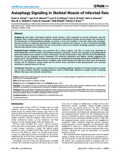
| dc.description.abstract | Background:
Heart failure (HF)-induced skeletal muscle atrophy is often associated to exercise intolerance and poor prognosis. Better understanding of the molecular mechanisms underlying HF-induced muscle atrophy may contribute to the development of pharmacological strategies to prevent or treat such condition. It has been shown that autophagy-lysosome system is an important mechanism for maintenance of muscle mass. However, its role in HF-induced myopathy has not been addressed yet. Therefore, the aim of the present study was to evaluate autophagy signaling in myocardial infarction (MI)-induced muscle atrophy in rats.
Methods/Principal Findings:
Wistar rats underwent MI or Sham surgeries, and after 12 weeks were submitted to echocardiography, exercise tolerance and histology evaluations. Cathepsin L activity and expression of autophagy-related genes and proteins were assessed in soleus and plantaris muscles by fluorimetric assay, qRT-PCR and immunoblotting, respectively. MI rats displayed exercise intolerance, left ventricular dysfunction and dilation, thereby suggesting the presence of HF. The key findings of the present study were: a) upregulation of autophagy-related genes (GABARAPL1, ATG7, BNIP3, CTSL1 and LAMP2) was observed only in plantaris while muscle atrophy was observed in both soleus and plantaris muscles, and b) Cathepsin L activity, Bnip3 and Fis1 protein levels, and levels of lipid hydroperoxides were increased specifically in plantaris muscle of MI rats.
Conclusions:
Altogether our results provide evidence for autophagy signaling regulation in HF-induced plantaris atrophy but not soleus atrophy. Therefore, autophagy-lysosome system is differentially regulated in atrophic muscles comprising different fiber-types and metabolic characteristics. | nb_NO |
| dc.language.iso | eng | nb_NO |
| dc.publisher | Public Library of Science | nb_NO |
| dc.title | Autophagy signaling in skeletal muscle of infarcted rats | nb_NO |
| dc.type | Journal article | nb_NO |
| dc.type | Peer reviewed | en_GB |
| dc.date.updated | 2015-11-24T12:46:33Z | |
| dc.source.volume | 9 | nb_NO |
| dc.source.journal | PLoS ONE | nb_NO |
| dc.source.issue | 1 | nb_NO |
| dc.identifier.doi | 10.1371/journal.pone.0085820 | |
| dc.identifier.cristin | 1128491 | |
| dc.description.localcode | © 2014 Jannig et al. This is an open-access article distributed under the terms of the Creative Commons Attribution License, which permits unrestricted use, distribution, and reproduction in any medium, provided the original author and source are credited. | nb_NO |