| dc.contributor.advisor | Vannesjo, Johanna | |
| dc.contributor.advisor | Beghini, Laura | |
| dc.contributor.author | Ponsi, Brunnhilde | |
| dc.date.accessioned | 2023-08-29T17:22:24Z | |
| dc.date.available | 2023-08-29T17:22:24Z | |
| dc.date.issued | 2023 | |
| dc.identifier | no.ntnu:inspera:146341341:93722241 | |
| dc.identifier.uri | https://hdl.handle.net/11250/3086283 | |
| dc.description.abstract | Bakgrunn: Magnetisk resonanstomografi (MR) er en effektiv avbildningsteknikk som gir overlegen kontrast i bløtvev sammenlignet med andre teknikker som brukes i klinisk praksis. Nye metoder innen MR krever hyppig testing, og særlig tidlig i utviklingsprosessen blir denne testingen vanligvis gjennomført ved skanning av homogene fantomer. Disse fantomene fanger imidlertid ikke opp utfordringene som oppstår in vivo. For eksempel kan forskjeller i magnetisk susceptibilitet på tvers av vev in vivo føre til feltinhomogeniteter, som videre kan forårsake signaltap i bildene. Dette gjelder spesielt for ryggmargsavbildning ved 7T, hvor data er sterkt påvirket av B0 feltinhomogenitet, grunnet susceptibilitetsforskjeller mellom ryggvirvlene og det omkringliggende vevet. «Slice-wise shimming»-teknikker har blitt foreslått for å redusere signalforvrengning og signaltap, men disse fjerner vanligvis ikke framtredende signalartefakter.
Et antropomorft MR-fantom av den menneskelige cervicalcolumna som etterligner den statiske B0 feltfordelingen, kan deretter brukes til å optimalisere avanserte shimming- og innhentingsmetoder for 7T MR-avbildning av ryggmargen.
Metoder: En første fantomprototype bestående av 3D-printede ryggvirvler C3 til C5 ble bygget og plassert i en sfærisk beholder. Ulike ingredienser og produksjonsmaterialer ble testet for å finjustere følsomhetsforskjellen mellom ryggvirvlene og løsningen, i tillegg til løsningens relaksasjonstid T2 star. Deretter ble en mer avansert fantomversjon utviklet, bestående av et 3D-printet fantomskall i form av et menneskehode og thorax med ryggvirvler fra C1 til C7.
Feltkart og gradient ekko (GRE) multiekkosekvenser av fantomene ble kartlagt ved 7T på et Siemens MAGNETOM Terra System.
Resultater: Det ble observert en omtrentlig samsvarelse mellom fantomkomponentene, in-vivo følsomhetsforskjellen mellom ryggvirvlene og omkringliggende vev, og ryggmargens T2 star verdi. Observerte feltkart av fantomet ble sammenlignet med feltsimuleringer, der disse presenterte lignende trekk med hensyn til egen feltforvrengning. Fantomet viste også et rommessig periodisk mønster av signaltap rundt de intervertebrale kryssene i multi-ekko GRE-bildene, i likhet med det som ofte observeres i in vivo-data. Signaltapet oppsto i områder med høyere feltinhomogenitet.
Konklusjon: Dette fantomet viser at det er mulig å 3D-printe et tilpasningsdyktig, ikke-toksisk antropomorft fantom som nøyaktig gjengir B0-feltmønstre fra ryggraden og kan benyttes til å teste nye anskaffelsesstrategier og etterbehandlingsstrategier for å løse den vedvarende utfordringen med statisk B0-feltforvrengning i ryggmargsavbildning. | |
| dc.description.abstract | Background: Magnetic resonance imaging (MRI) is a powerful imaging technique that offers superior soft tissue contrast compared to other techniques used in the clinical practice. Innovation in MRI requires frequent testing, commonly done scanning homogeneous phantoms, especially at an early stage. These phantoms, however, fail to represent the challenges encountered in vivo. For instance, magnetic susceptibility differences across tissues lead in vivo to field inhomogeneities causing signal loss in the images. This is particularly true for spinal cord imaging at 7T where data are heavily affected by B0 field inhomogeneity, due to susceptibility difference between the vertebrae and the surrounding tissues. Slice-wise shimming techniques have been proposed to reduce the distortion and signal drop-out but substantial artifacts typically persist.
An anthropomorphic MRI phantom of the human cervical spine mimicking the static B0 field distribution could then be used to optimize advanced shimming and acquisition techniques for 7T spinal cord MRI.
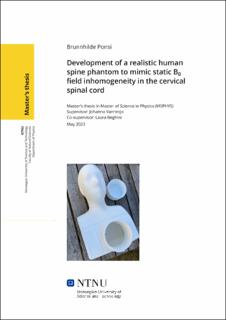
Methods: An initial phantom prototype consisting of 3D printed vertebrae C3-to-C5 in a spherical container was built. Various ingredients and printing materials were tested to tune the susceptibility difference between the vertebrae and the solution, along with the relaxation time T2 star of the solution. Subsequently, a more advanced phantom version was developed, consisting of a 3D printed phantom shell in the shape of a human head and thorax containing vertebrae from C1 to C7.
Field maps and gradient echo (GRE) multi-echo sequences of the phantoms were acquired at 7T on a Siemens MAGNETOM Terra System.
Results: The phantom components approximately matched the in vivo susceptibility difference between the vertebrae and surrounding tissues, as well as the spinal cord T2 star value. Measured field maps of the phantom were compared with field simulations, showing similar features in the field distortion. The phantom also exhibited a spatially periodic pattern of signal drop-out around the intervertebral junctions in multi-echo GRE images, similar to what is commonly observed in in vivo data. The signal drop-out arises in regions with higher field inhomogeneities.
Conclusion: This phantom demonstrates the feasibility of 3D printing an adaptable, non-toxic anthropomorphic phantom that accurately reproduces B0 field patterns from the spine and may serve to test new acquisition strategies and post-processing strategies to address the persistent challenge of static B0 field distortion in spinal cord imaging. | |
| dc.language | eng | |
| dc.publisher | NTNU | |
| dc.title | Development of a realistic human spine phantom to mimic static B0 field inhomogeneity in the cervical spinal cord | |
| dc.type | Master thesis | |
