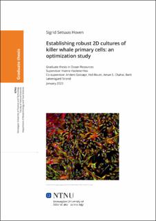
Establishing robust 2D cultures of killer whale primary cells: an optimization study
Master thesis
Permanent lenke
https://hdl.handle.net/11250/3078439Utgivelsesdato
2023Metadata
Vis full innførselSamlinger
Sammendrag
Norske spekkhoggere (Orcinus orca) er som følge av deres trofiske nivå og føde blant de mest kontaminerte marine pattedyrene i verden. Akkumulering av persistente organiske forbindelser (POPs) kan føre til forstyrrelser av biokjemiske prosesser på cellulært nivå som kan ha alvorlige innvirkninger på populasjonene. I løpet av det siste tiåret er det lagt økt innsats i å etablere in vitro modellsystem for marine pattedyr med den hensikt å kunne studere hvilke konsenkvenser miljøgiftene kan ha på dyrene. Målet med denne masteroppgaven var å etablere en stabil 2D cellekultur med primære fibroblaster derivert fra vevsprøver tatt av norske fritt-levende spekkhogger gjennom å optimalisere kultiveringsforholdene. Primære dermale celler med fibroblast-typisk morfologi ble isolert og identifisert som fibroblaster via immunohistokjemi med primære vimentin antistoffer. Cellemorfologien ble også karakterisert med DAPI og Phalloidin farging av cellekjernen og intermediære aktinfilamenter. De isolerte spekkhogger fibroblastene ble dyrket i ulike kultiveringsforhold, med Dulbecco′s Modified Eagle′s Medium/Nutrient Mixture F-12 medium med høy eller lav glukosekonsentrasjon and utvalgte supplementeringer. Vi testet ulike konsentrasjoner av fibroblast vekstfaktor 2, fetalt kalveserum eller hesteserum, ikke-essensielle aminosyrer, askorbinsyre og 2-mercaptoetanol antioksidanter og gelatin eller laminin overflate behandlinger for å undersøke effekten av de ulike parameterene alene og i kombinasjon på cellevekst og cellemetabolisme. Celleproliferasjon og metabolise ble målt kvantitativt ved celletellinger og metabolittanalyser av glukose konsum og laktatakkumulering. Vi støtte på utfordringer, som begrenset tilgang på vevsmateriale og celler, saktevoksende celler og lav replikativ kapasitet og tidlig irreversibel celledvale (senescence), som er vanlig med primære hvalcellekulturer. Senescente celler ble bekreftet med β-galaktosidase senescent analyse. Friske, prolifererende fibroblaster ble også idenfisert via immunohistokjemi med primære Ki-67 antistoffer. Tilsats av fibroblast vekstfaktor resulterte i betydelig økning i proliferasjon ved 1 ng/mL konsentrasjon og den optimale konsentrasjonen var 2 ng/mL. De beste parameterene for cellevekst ble kobinert og bestod av 2 ng/mL fibroblast vekstfaktor, 15 % fetalt kalveserum, 5 % hesteserum og 0.1 mM 2-mercaptoetanol. Med det optimaliserte mediet oppnådde vi 20 passasjer med primære fibroblaster fra spekkhoggere uten å se tegn til reduksjon i vekstrate. Populasjonsdoblingstiden ble beregnet til å være 40 timer. Den etablerte cellekulturen ble videre brukt til å måle cytotoksisk effekt av en miks med de 10 vanligste miljøgiftene som er målt i spekket hos norske spekkhoggere. Vi fant en dødelighet på 25 % for den høyeste eksponerte konsentrasjonen på 202.1 μg/mL, tilsvarene 50X den målte konsentrasjonen i spekket. Flere POPs, blant annet polyklorerte bifenyler og p,p’-DDT er kjent for å forstyrre cellulære mekanismer ved å binde seg til ulike reseptorer som aktivere og endrer genuttrykk i cellene. Videre forskning vil være nødvendig for å skaffe mer kunnskap om hvordan spekkhoggere påvirkes av miljøgifter på genetisk og biokjemisk nivå. Due to their trophic level and sources of prey, top predator Norwegian killer whales (Orcinus orca) are among the most contaminated marine species throughout the world. Accumulation of persistant organic pollutants (POPs) may have adverse effects on killer whale populations, by disrupting vital biochemical processes. Over the last decade, increased effort has been put into establishing in vitro model systems for marine mammals to assess the potential hazardous effects inflicted by environmental toxicants. The aim of this thesis was to establish a robust 2D cell culture with primary fibroblast cells derived from Norwegian killer whale skin tissue samples from free-ranging specimens by optimizing the cultivation conditions. Primary dermal cells exhibiting the typical spindle-shape of fibroblasts were isolated and identified as fibroblasts via primary vimentin antibody immunolabellering. Cell morphology was characterized with DAPI and phalloidin staining. The isolated killer whale fibroblasts were cultivated in various cultivation conditions, using Dulbecco′s Modified Eagle′s Medium/Nutrient Mixture F-12 media with high or low concentration of glucose and varying selected supplementations. We tested different concentrations of fibroblast growth factor 2, fetal bovine serum or horse serum, non-essential amino acids, ascorbic acid and 2-Mercaptoethanol antioxidants, and gelatin or laminin surface coatings, to investigate the effect on cell growth and cell metabolism by the different parameters indiviually and in combinations. Cell proliferation and metabolism was measured by cell counting and metabolite analysis of glucose consumption and lactate accumulation. Common challenges associated with cetacean cell cultures were encountered, including limitation of cell sources, slow cell proliferation rates and senescence, which was confirmed in killer whale fibroblasts with the β-galactosidase senescence assay. Immunhistochemistry was successfully applied using primary Ki-67 antibody to identify proliferating fibroblast cells. FGF supplementation increased proliferation significantly at 1 ng/mL and an optimal concentration of 2 ng/mL. The optimized media composed of bFGF 2 ng/ml supplemented with 15 % FBS and 5 % horse serum and 2-Mercaptoethanol antioxidant, maintained a killer whale cell culture for 20 passages with a cell population doubling time of 40 hours without showing signs of reduced proliferation or senescence. The established cell culture was further used to assess cytotoxic effects of a cocktail which contained the most common POPs found in killer whale blubber after 48 hours exposure. A 25% loss in cell metabolism was detected in killer whale fibroblasts at the highest exposure concentration of 50X (202.1 μg/mL) the measured concentration in killer whale blubber with the CellTiter-Glo assay, while no cytotoxicity was detected with LDH release assay. Several POPs, including PCBs and p,p’-DDT, are known to bind to several receptors activating and changing gene expression and by so disrupting cellular mechanism. Further research is necessary to increase our knowledge about how killer whales are affected by environmental toxicants on genetic and biochemical level.
