| dc.contributor.author | Nestaas, Sverre | |
| dc.contributor.author | STENSÆTH, KNUT-HAAKON | |
| dc.contributor.author | Rosseland, Vigdis | |
| dc.contributor.author | Kramer-Johansen, Jo | |
| dc.date.accessioned | 2020-03-25T13:14:48Z | |
| dc.date.available | 2020-03-25T13:14:48Z | |
| dc.date.created | 2016-07-15T15:16:42Z | |
| dc.date.issued | 2016 | |
| dc.identifier.citation | Scandinavian Journal of Trauma, Resuscitation and Emergency Medicine. 2016, 24 (54), . | en_US |
| dc.identifier.issn | 1757-7241 | |
| dc.identifier.uri | https://hdl.handle.net/11250/2648609 | |
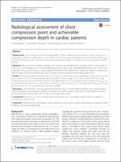
| dc.description.abstract | Background
Using magnetic resonance imaging (MRI) to relate cardiovascular structures to surface anatomy in a population relevant to cardiac arrest victims, relate the external thoracic anterior-posterior (AP) diameter (APEXTERNAL) and blood-filled structures to recommended chest compression depths, and define an optimal compression point (OCP).
Methods
MRI axial scans of referred patients were analysed. We defined origo as the skin surface of the centre of sternum in the internipple line. The blood-filled structures beneath origo were identified and the sum of their inner diameters (APBLOOD) and APEXTERNAL were measured. We defined OCP based on the image with maximum compressible left and right ventricle and where LVOT was not present. We measured the distance from origo to OCP.
Results
Consecutive patients, mean (SD), age 52 (17) years, 110 (76 %) males, were categorized: cardiac disease (n = 74), aortic disease (n = 13), no findings/study patient (included in another study) (n = 57). The structure LVOT/aortic valve (AV)/aortic root was present in 46 % of patients with cardiac disease vs. 19 % of patients with no findings. APEXTERNAL for males and females was 25 (2) cm and 22 (2) cm, and APBLOOD 6.5 cm (2) and 4.7 cm (2), respectively. Distance from origo to OCP was 32 (11) mm to the left and 16 (21) mm caudally.
Discussion
LVOT/AV/aortic root was present beneath the origo in almost half the patients with cardiac disease. Recommended chest compression depths exceeded the anterior-posterior diameter of blood-filled structures in more than half of the females. OCP was found 3 cm left of the origo.
Conclusions
Based on our study, individualized compression point and depth could be further studied in a prospective, clinical study. | en_US |
| dc.language.iso | eng | en_US |
| dc.publisher | BioMed Central | en_US |
| dc.rights | Navngivelse 4.0 Internasjonal | * |
| dc.rights.uri | http://creativecommons.org/licenses/by/4.0/deed.no | * |
| dc.title | Radiological assessment of chest compression point and achievable compression depth in cardiac patients | en_US |
| dc.type | Peer reviewed | en_US |
| dc.type | Journal article | en_US |
| dc.description.version | publishedVersion | en_US |
| dc.source.pagenumber | 8 | en_US |
| dc.source.volume | 24 | en_US |
| dc.source.journal | Scandinavian Journal of Trauma, Resuscitation and Emergency Medicine | en_US |
| dc.source.issue | 54 | en_US |
| dc.identifier.doi | 10.1186/s13049-016-0245-0 | |
| dc.identifier.cristin | 1368218 | |
| dc.description.localcode | Open Access This article is distributed under the terms of the Creative Commons Attribution 4.0 International License (http://creativecommons.org/licenses/by/4.0/), which permits unrestricted use, distribution, and reproduction in any medium, provided you give appropriate credit to the original author(s) and the source, provide a link to the Creative Commons license, and indicate if changes were made. The Creative Commons Public Domain Dedication waiver (http://creativecommons.org/publicdomain/zero/1.0/) applies to the data made available in this article, unless otherwise stated | en_US |
| cristin.ispublished | true | |
| cristin.fulltext | original | |
| cristin.qualitycode | 1 | |

