| dc.contributor.author | Maas, Marnix C | |
| dc.contributor.author | Litjens, Geert | |
| dc.contributor.author | Wright, A. J. | |
| dc.contributor.author | Attenberger, UI | |
| dc.contributor.author | Haider, MA | |
| dc.contributor.author | Helbich, Thomas H. | |
| dc.contributor.author | Kiefer, B | |
| dc.contributor.author | Macura, KJ | |
| dc.contributor.author | Margolis, David Joel | |
| dc.contributor.author | Padhani, AR | |
| dc.contributor.author | Selnæs, Kirsten Margrete | |
| dc.contributor.author | Villeirs, GM | |
| dc.contributor.author | Futterer, Jürgen J. | |
| dc.contributor.author | Scheenen, Tom WJ | |
| dc.date.accessioned | 2020-03-05T08:15:42Z | |
| dc.date.available | 2020-03-05T08:15:42Z | |
| dc.date.created | 2019-04-08T10:54:47Z | |
| dc.date.issued | 2019 | |
| dc.identifier.citation | Investigative Radiology. 2019, 54 (7), 437-447. | nb_NO |
| dc.identifier.issn | 0020-9996 | |
| dc.identifier.uri | http://hdl.handle.net/11250/2645378 | |
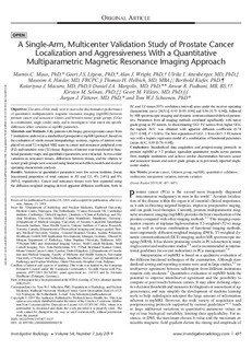
| dc.description.abstract | Objectives: The aims of this study were to assess the discriminative performance of quantitative multiparametric magnetic resonance imaging (mpMRI) between prostate cancer and noncancer tissues and between tumor grade groups (GGs) in a multicenter, single-vendor study, and to investigate to what extent site-specific differences affect variations in mpMRI parameters.
Materials and Methods: Fifty patients with biopsy-proven prostate cancer from 5 institutions underwent a standardized preoperative mpMRI protocol. Based on the evaluation of whole-mount histopathology sections, regions of interest were placed on axial T2-weighed MRI scans in cancer and noncancer peripheral zone (PZ) and transition zone (TZ) tissue. Regions of interest were transferred to functional parameter maps, and quantitative parameters were extracted. Across-center variations in noncancer tissues, differences between tissues, and the relation to cancer grade groups were assessed using linear mixed-effects models and receiver operating characteristic analyses.
Results: Variations in quantitative parameters were low across institutes (mean [maximum] proportion of total variance in PZ and TZ, 4% [14%] and 8% [46%], respectively). Cancer and noncancer tissues were best separated using the diffusion-weighted imaging-derived apparent diffusion coefficient, both in PZ and TZ (mean [95% confidence interval] areas under the receiver operating characteristic curve [AUCs]; 0.93 [0.89–0.96] and 0.86 [0.75–0.94]), followed by MR spectroscopic imaging and dynamic contrast-enhanced-derived parameters. Parameters from all imaging methods correlated significantly with tumor grade group in PZ tumors. In discriminating GG1 PZ tumors from higher GGs, the highest AUC was obtained with apparent diffusion coefficient (0.74 [0.57–0.90], P < 0.001). The best separation of GG1–2 from GG3–5 PZ tumors was with a logistic regression model of a combination of functional parameters (mean AUC, 0.89 [0.78–0.98]).
Conclusions: Standardized data acquisition and postprocessing protocols in prostate mpMRI at 3 T produce equivalent quantitative results across patients from multiple institutions and achieve similar discrimination between cancer and noncancer tissues and cancer grade groups as in previously reported singlecenter studies. | nb_NO |
| dc.language.iso | eng | nb_NO |
| dc.publisher | Lippincott, Williams & Wilkins | nb_NO |
| dc.rights | Attribution-NonCommercial-NoDerivatives 4.0 Internasjonal | * |
| dc.rights.uri | http://creativecommons.org/licenses/by-nc-nd/4.0/deed.no | * |
| dc.title | A Single-Arm, Multicenter Validation Study of Prostate Cancer Localization and Aggressiveness With a Quantitative Multiparametric Magnetic Resonance Imaging Approach. | nb_NO |
| dc.type | Journal article | nb_NO |
| dc.type | Peer reviewed | nb_NO |
| dc.description.version | publishedVersion | nb_NO |
| dc.source.pagenumber | 437-447 | nb_NO |
| dc.source.volume | 54 | nb_NO |
| dc.source.journal | Investigative Radiology | nb_NO |
| dc.source.issue | 7 | nb_NO |
| dc.identifier.doi | 10.1097/RLI.0000000000000558 | |
| dc.identifier.cristin | 1690782 | |
| dc.description.localcode | © 2019 The Author(s). Published by Wolters Kluwer Health, Inc. This is an open-access article distributed under the terms of the Creative Commons Attribution-Non Commercial-No Derivatives License 4.0 (CC-BY-NC-ND), where it is permissible to download and share the work provided it is properly cited. The work cannot be changed in any way or used commercially without permission from the journal. | nb_NO |
| cristin.unitcode | 194,65,25,0 | |
| cristin.unitname | Institutt for sirkulasjon og bildediagnostikk | |
| cristin.ispublished | true | |
| cristin.fulltext | original | |
| cristin.qualitycode | 2 | |

