Brain Organization of Apolygus lucorum: A Hemipteran Species With Prominent Antennal Lobes
| dc.contributor.author | Xie, Gui-Ying | |
| dc.contributor.author | Ma, Bai-Wei | |
| dc.contributor.author | Liu, Xiao-Lan | |
| dc.contributor.author | Chang, Ya-Jun | |
| dc.contributor.author | Chen, Wen-Bo | |
| dc.contributor.author | Li, Guo-Ping | |
| dc.contributor.author | Feng, Hong-Qiang | |
| dc.contributor.author | Zhang, Yong-Jun | |
| dc.contributor.author | Berg, Bente Gunnveig | |
| dc.contributor.author | Zhao, Xin-Cheng | |
| dc.date.accessioned | 2020-02-10T12:09:16Z | |
| dc.date.available | 2020-02-10T12:09:16Z | |
| dc.date.created | 2019-09-17T14:40:18Z | |
| dc.date.issued | 2019 | |
| dc.identifier.citation | Frontiers in Neuroanatomy. 2019, 13 (70), 1-13. | nb_NO |
| dc.identifier.issn | 1662-5129 | |
| dc.identifier.uri | http://hdl.handle.net/11250/2640694 | |
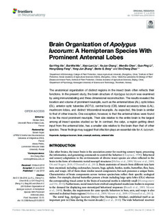
| dc.description.abstract | The anatomical organization of distinct regions in the insect brain often reflects their functions. In the present study, the brain structure of Apolygus lucorum was examined by using immunolabeling and three-dimensional reconstruction. The results revealed the location and volume of prominent neuropils, such as the antennal lobes (AL), optic lobes (OL), anterior optic tubercles (AOTU), central body (CB), lateral accessory lobes (LAL), mushroom lobes, and distinct tritocerebral neuropils. As expected, this brain is similar to that of other insects. One exception, however, is that the antennal lobes were found to be the most prominent neuropils. Their size relative to the entire brain is the largest among all insect species studied so far. In contrast, the calyx, a region getting direct input from the antennal lobe, has a smaller size relative to the brain than that of other species. These findings may suggest that olfaction plays an essential role for A. lucorum. | nb_NO |
| dc.language.iso | eng | nb_NO |
| dc.publisher | Frontiers Media | nb_NO |
| dc.rights | Navngivelse 4.0 Internasjonal | * |
| dc.rights.uri | http://creativecommons.org/licenses/by/4.0/deed.no | * |
| dc.title | Brain Organization of Apolygus lucorum: A Hemipteran Species With Prominent Antennal Lobes | nb_NO |
| dc.type | Journal article | nb_NO |
| dc.type | Peer reviewed | nb_NO |
| dc.description.version | publishedVersion | nb_NO |
| dc.source.pagenumber | 1-13 | nb_NO |
| dc.source.volume | 13 | nb_NO |
| dc.source.journal | Frontiers in Neuroanatomy | nb_NO |
| dc.source.issue | 70 | nb_NO |
| dc.identifier.doi | 10.3389/fnana.2019.00070 | |
| dc.identifier.cristin | 1725736 | |
| dc.description.localcode | Copyright © 2019 Xie, Ma, Liu, Chang, Chen, Li, Feng, Zhang, Berg and Zhao. This is an open-access article distributed under the terms of the Creative Commons Attribution License (CC BY). The use, distribution or reproduction in other forums is permitted, provided the original author(s) and the copyright owner(s) are credited and that the original publication in this journal is cited, in accordance with accepted academic practice. No use, distribution or reproduction is permitted which does not comply with these terms. | nb_NO |
| cristin.unitcode | 194,67,40,0 | |
| cristin.unitname | Institutt for psykologi | |
| cristin.ispublished | true | |
| cristin.fulltext | original | |
| cristin.qualitycode | 1 |
Tilhørende fil(er)
Denne innførselen finnes i følgende samling(er)
-
Institutt for psykologi [2883]
-
Publikasjoner fra CRIStin - NTNU [37177]

Med mindre annet er angitt, så er denne innførselen lisensiert som Navngivelse 4.0 Internasjonal