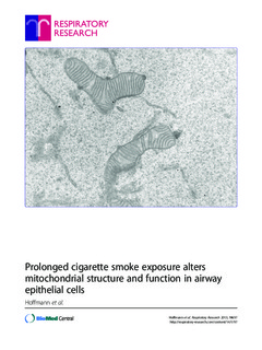
Prolonged cigarette smoke exposure alters mitochondrial structure and function in airway epithelial cells
| dc.contributor.author | Hoffmann, RF | |
| dc.contributor.author | Zarrintan, S | |
| dc.contributor.author | Brandenburg, SM | |
| dc.contributor.author | Kol, A | |
| dc.contributor.author | de Bruin, HG | |
| dc.contributor.author | Jafari, S | |
| dc.contributor.author | Dijk, F | |
| dc.contributor.author | Kalicharan, D | |
| dc.contributor.author | Kelders, M | |
| dc.contributor.author | Gosker, HR | |
| dc.contributor.author | ten Hacken, NHT | |
| dc.contributor.author | Van der Want, Johannes | |
| dc.contributor.author | van Oosterhout, AJM | |
| dc.contributor.author | Heijink, IH | |
| dc.date.accessioned | 2019-10-28T07:27:11Z | |
| dc.date.available | 2019-10-28T07:27:11Z | |
| dc.date.created | 2014-01-23T08:52:48Z | |
| dc.date.issued | 2013 | |
| dc.identifier.citation | Respiratory Research. 2013, 14 . | nb_NO |
| dc.identifier.issn | 1465-9921 | |
| dc.identifier.uri | http://hdl.handle.net/11250/2624740 | |
| dc.description.abstract | Background Cigarette smoking is the major risk factor for COPD, leading to chronic airway inflammation. We hypothesized that cigarette smoke induces structural and functional changes of airway epithelial mitochondria, with important implications for lung inflammation and COPD pathogenesis. Methods We studied changes in mitochondrial morphology and in expression of markers for mitochondrial capacity, damage/biogenesis and fission/fusion in the human bronchial epithelial cell line BEAS-2B upon 6-months from ex-smoking COPD GOLD stage IV patients to age-matched smoking and never-smoking controls. Results We observed that long-term CSE exposure induces robust changes in mitochondrial structure, including fragmentation, branching and quantity of cristae. The majority of these changes were persistent upon CSE depletion. Furthermore, long-term CSE exposure significantly increased the expression of specific fission/fusion markers (Fis1, Mfn1, Mfn2, Drp1 and Opa1), oxidative phosphorylation (OXPHOS) proteins (Complex II, III and V), and oxidative stress (Mn-SOD) markers. These changes were accompanied by increased levels of the pro-inflammatory mediators IL-6, IL-8, and IL-1β. Importantly, COPD primary bronchial epithelial cells (PBECs) displayed similar changes in mitochondrial morphology as observed in long-term CSE-exposure BEAS-2B cells. Moreover, expression of specific OXPHOS proteins was higher in PBECs from COPD patients than control smokers, as was the expression of mitochondrial stress marker PINK1. Conclusion The observed mitochondrial changes in COPD epithelium are potentially the consequence of long-term exposure to cigarette smoke, leading to impaired mitochondrial function and may play a role in the pathogenesis of COPD. | nb_NO |
| dc.language.iso | eng | nb_NO |
| dc.publisher | BMC (part of Springer Nature) | nb_NO |
| dc.rights | Navngivelse 4.0 Internasjonal | * |
| dc.rights.uri | http://creativecommons.org/licenses/by/4.0/deed.no | * |
| dc.title | Prolonged cigarette smoke exposure alters mitochondrial structure and function in airway epithelial cells | nb_NO |
| dc.type | Journal article | nb_NO |
| dc.type | Peer reviewed | nb_NO |
| dc.description.version | publishedVersion | nb_NO |
| dc.source.pagenumber | 12 | nb_NO |
| dc.source.volume | 14 | nb_NO |
| dc.source.journal | Respiratory Research | nb_NO |
| dc.identifier.doi | 10.1186/1465-9921-14-97 | |
| dc.identifier.cristin | 1097822 | |
| dc.description.localcode | © 2013 Hoffmann et al.; licensee BioMed Central Ltd. This is an Open Access article distributed under the terms of the Creative Commons Attribution License (http://creativecommons.org/licenses/by/2.0), which permits unrestricted use, distribution, and reproduction in any medium, provided the original work is properly cited. | nb_NO |
| cristin.unitcode | 194,65,15,0 | |
| cristin.unitname | Institutt for klinisk og molekylær medisin | |
| cristin.ispublished | true | |
| cristin.fulltext | original | |
| cristin.qualitycode | 1 |
Files in this item
This item appears in the following Collection(s)

Except where otherwise noted, this item's license is described as Navngivelse 4.0 Internasjonal