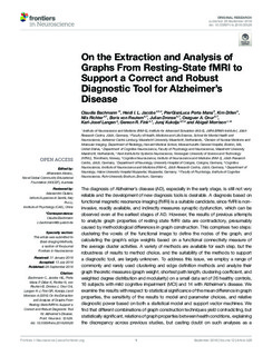
On the Extraction and Analysis of Graphs From Resting-State fMRI to Support a Correct and Robust Diagnostic Tool for Alzheimer's Disease
| dc.contributor.author | Bachmann, Claudia | |
| dc.contributor.author | Jacobs, Heidi I. L. | |
| dc.contributor.author | Mana, Piero | |
| dc.contributor.author | Dillen, Kim | |
| dc.contributor.author | Richter, Nils | |
| dc.contributor.author | von Reutern, Boris | |
| dc.contributor.author | Dronse, Julian | |
| dc.contributor.author | Onur, Oezguer A. | |
| dc.contributor.author | Langen, Karl-Josef | |
| dc.contributor.author | Fink, Gereon R. | |
| dc.contributor.author | Kukolja, Juraj | |
| dc.contributor.author | Morrison, Abigail | |
| dc.date.accessioned | 2019-09-19T08:16:06Z | |
| dc.date.available | 2019-09-19T08:16:06Z | |
| dc.date.created | 2019-01-18T14:00:53Z | |
| dc.date.issued | 2018 | |
| dc.identifier.citation | Frontiers in Neuroscience. 2018, 12 | nb_NO |
| dc.identifier.issn | 1662-4548 | |
| dc.identifier.uri | http://hdl.handle.net/11250/2617580 | |
| dc.description.abstract | The diagnosis of Alzheimer's disease (AD), especially in the early stage, is still not very reliable and the development of new diagnosis tools is desirable. A diagnosis based on functional magnetic resonance imaging (fMRI) is a suitable candidate, since fMRI is non-invasive, readily available, and indirectly measures synaptic dysfunction, which can be observed even at the earliest stages of AD. However, the results of previous attempts to analyze graph properties of resting state fMRI data are contradictory, presumably caused by methodological differences in graph construction. This comprises two steps: clustering the voxels of the functional image to define the nodes of the graph, and calculating the graph's edge weights based on a functional connectivity measure of the average cluster activities. A variety of methods are available for each step, but the robustness of results to method choice, and the suitability of the methods to support a diagnostic tool, are largely unknown. To address this issue, we employ a range of commonly and rarely used clustering and edge definition methods and analyze their graph theoretic measures (graph weight, shortest path length, clustering coefficient, and weighted degree distribution and modularity) on a small data set of 26 healthy controls, 16 subjects with mild cognitive impairment (MCI) and 14 with Alzheimer's disease. We examine the results with respect to statistical significance of the mean difference in graph properties, the sensitivity of the results to model and parameter choices, and relative diagnostic power based on both a statistical model and support vector machines. We find that different combinations of graph construction techniques yield contradicting, but statistically significant, relations of graph properties between health conditions, explaining the discrepancy across previous studies, but casting doubt on such analyses as a method to gain insight into disease effects. The production of significant differences in mean graph properties turns out not to be a good predictor of future diagnostic capacity. Highest predictive power, expressed by largest negative surprise values, are achieved for both atlas-driven and data-driven clustering (Ward clustering), as long as graphs are small and clusters large, in combination with edge definitions based on correlations and mutual information transfer. | nb_NO |
| dc.language.iso | eng | nb_NO |
| dc.publisher | Frontiers Media | nb_NO |
| dc.relation.uri | https://doi.org/10.3389/fnins.2018.00528 | |
| dc.rights | Navngivelse 4.0 Internasjonal | * |
| dc.rights.uri | http://creativecommons.org/licenses/by/4.0/deed.no | * |
| dc.title | On the Extraction and Analysis of Graphs From Resting-State fMRI to Support a Correct and Robust Diagnostic Tool for Alzheimer's Disease | nb_NO |
| dc.type | Journal article | nb_NO |
| dc.type | Peer reviewed | nb_NO |
| dc.description.version | publishedVersion | nb_NO |
| dc.source.volume | 12 | nb_NO |
| dc.source.journal | Frontiers in Neuroscience | nb_NO |
| dc.identifier.doi | 10.3389/fnins.2018.00528 | |
| dc.identifier.cristin | 1660357 | |
| dc.description.localcode | Copyright © 2018 Bachmann, Jacobs, Porta Mana, Dillen, Richter, von Reutern, Dronse, Onur, Langen, Fink, Kukolja and Morrison. This is an open-access article distributed under the terms of the Creative Commons Attribution License (CC BY). The use, distribution or reproduction in other forums is permitted, provided the original author(s) and the copyright owner(s) are credited and that the original publication in this journal is cited, in accordance with accepted academic practice. No use, distribution or reproduction is permitted which does not comply with these terms. | nb_NO |
| cristin.unitcode | 194,65,60,0 | |
| cristin.unitname | Kavliinstitutt for nevrovitenskap | |
| cristin.ispublished | true | |
| cristin.fulltext | original | |
| cristin.qualitycode | 1 |
Tilhørende fil(er)
Denne innførselen finnes i følgende samling(er)

Med mindre annet er angitt, så er denne innførselen lisensiert som Navngivelse 4.0 Internasjonal