| dc.contributor.author | Rusandu, Albertina | |
| dc.contributor.author | Ødegård, Asbjørn | |
| dc.contributor.author | Engh, Gabriele | |
| dc.contributor.author | Olerud, Hilde M | |
| dc.date.accessioned | 2019-02-12T14:31:55Z | |
| dc.date.available | 2019-02-12T14:31:55Z | |
| dc.date.created | 2018-11-09T12:31:25Z | |
| dc.date.issued | 2018 | |
| dc.identifier.issn | 1078-8174 | |
| dc.identifier.uri | http://hdl.handle.net/11250/2585072 | |
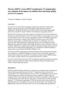
| dc.description.abstract | Introduction
Use of CT in the investigation of pulmonary embolism in radiosensitive patients such as pregnant and young female patients entails the need for protocol optimization. The aim of this study was to analyze the dose reduction and image quality achieved by using 80 kV instead of 100 kV in CT pulmonary angiography protocols.
Methods
80 examinations of non-obese patients were analyzed (40 consecutive patients for each protocol, equally distributed on two CT scanners). Objective image quality was assessed by measurements of HU values (average and standard deviation) in five ROIs in pulmonary arteries and calculations of signal-to-noise (SNR) and contrast-to-noise ratios (CNR). Subjective image quality was independently evaluated by two radiologists in terms of perceived noise, sharp reproduction of pulmonary arteries and overall diagnostic quality. Radiation dose parameters (CTDIvol, DLP, SSDE and effective dose) and effective risk were compared. Differences in radiation dose and objective measures of image quality for the two protocols were assessed using the independent t test; comparison of subjective grading of image quality was performed with the Mann–Whitney U test.
Results
Use of 80 kV significantly increased both arterial contrast enhancement and image noise. Differences in SNR and CNR between protocols were not statistically significant. Achieved dose reduction by using 80 kV was significant on both scanners (SSDE reduction 35% and 46%, p < 0.001; effective dose reduction 40% and 53%, p < 0.001).
Conclusion
Use of 80 kV protocols for CT examinations of pulmonary arteries in non-obese patients with bodyweight below 80 kg results in significant reduction of radiation doses without compromising image quality. | nb_NO |
| dc.language.iso | eng | nb_NO |
| dc.publisher | Elsevier | nb_NO |
| dc.rights | Attribution-NonCommercial-NoDerivatives 4.0 Internasjonal | * |
| dc.rights.uri | http://creativecommons.org/licenses/by-nc-nd/4.0/deed.no | * |
| dc.title | The use of 80 kV versus 100 kV in pulmonary CT angiography: An evaluation of the impact on radiation dose and image quality on two CT scanners | nb_NO |
| dc.title.alternative | The use of 80 kV versus 100 kV in pulmonary CT angiography: An evaluation of the impact on radiation dose and image quality on two CT scanners | nb_NO |
| dc.type | Journal article | nb_NO |
| dc.type | Peer reviewed | nb_NO |
| dc.description.version | acceptedVersion | nb_NO |
| dc.source.volume | 25 | nb_NO |
| dc.source.journal | Radiography | nb_NO |
| dc.source.issue | 1 | nb_NO |
| dc.identifier.doi | doi.org/10.1016/j.radi.2018.10.004 | |
| dc.identifier.cristin | 1628674 | |
| dc.description.localcode | © 2018. This is the authors’ accepted and refereed manuscript to the article. Locked until 8.1.2019 due to copyright restrictions. This manuscript version is made available under the CC-BY-NC-ND 4.0 license http://creativecommons.org/licenses/by-nc-nd/4.0/ | nb_NO |
| cristin.unitcode | 194,65,25,0 | |
| cristin.unitname | Institutt for sirkulasjon og bildediagnostikk | |
| cristin.ispublished | false | |
| cristin.fulltext | postprint | |
| cristin.qualitycode | 1 | |

