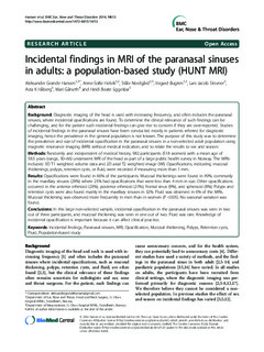
Incidental findings in MRI of the paranasal sinuses in adults: a population-based study (HUNT MRI)
| dc.contributor.author | Hansen, Aleksander Grande | |
| dc.contributor.author | Helvik, Anne-Sofie | |
| dc.contributor.author | Nordgård, Ståle | |
| dc.contributor.author | Bugten, Vegard | |
| dc.contributor.author | Stovner, Lars Jacob | |
| dc.contributor.author | Håberg, Asta | |
| dc.contributor.author | Gårseth, Mari | |
| dc.contributor.author | Eggesbø, Heidi Beate | |
| dc.date.accessioned | 2015-01-06T10:19:08Z | |
| dc.date.accessioned | 2016-04-20T11:34:54Z | |
| dc.date.available | 2015-01-06T10:19:08Z | |
| dc.date.available | 2016-04-20T11:34:54Z | |
| dc.date.issued | 2014 | |
| dc.identifier.citation | BMC Ear, Nose and Throat Disorders 2014, 14 | nb_NO |
| dc.identifier.issn | 1472-6815 | |
| dc.identifier.uri | http://hdl.handle.net/11250/2386495 | |
| dc.description.abstract | Background: Diagnostic imaging of the head is used with increasing frequency, and often includes the paranasal sinuses, where incidental opacifications are found. To determine the clinical relevance of such findings can be challenging, and for the patient such incidental findings can give rise to concern if they are over-reported. Studies of incidental findings in the paranasal sinuses have been conducted mostly in patients referred for diagnostic imaging, hence the prevalence in the general population is not known. The purpose of this study was to determine the prevalence and size of incidental opacification in the paranasal sinuses in a non-selected adult population using magnetic resonance imaging (MRI) without medical indication, and to relate the results to sex and season. Methods: Randomly and independent of medical history, 982 participants (518 women) with a mean age of 58.5 years (range, 50–66) underwent MRI of the head as part of a large public health survey in Norway. The MRIs included 3D T1 weighted volume data and 2D axial T2 weighted image (WI). Opacifications, indicating mucosal thickenings, polyps, retention cysts, or fluid, were recorded if measuring more than 1 mm. Results: Opacifications were found in 66% of the participants. Mucosal thickenings were found in 49%, commonly in the maxillary sinuses (29%) where 25% had opacifications that were less than 4 mm in size. Other opacifications occurred in the anterior ethmoid (23%), posterior ethmoid (21%), frontal sinus (9%), and sphenoid (8%). Polyps and retention cysts were also found mainly in the maxillary sinuses in 32%. Fluid was observed in 6% of the MRIs. Mucosal thickening was observed more frequently in men than in women (P <0.05). No seasonal variation was found. Conclusions: In this large non-selected sample, incidental opacification in the paranasal sinuses was seen in two out of three participants, and mucosal thickening was seen in one out of two. Fluid was rare. Knowledge of incidental opacification is important because it can affect clinical practice. | nb_NO |
| dc.language.iso | eng | nb_NO |
| dc.publisher | BioMed Central | nb_NO |
| dc.rights | Navngivelse 3.0 Norge | * |
| dc.rights.uri | http://creativecommons.org/licenses/by/3.0/no/ | * |
| dc.title | Incidental findings in MRI of the paranasal sinuses in adults: a population-based study (HUNT MRI) | nb_NO |
| dc.type | Journal article | nb_NO |
| dc.type | Peer reviewed | nb_NO |
| dc.date.updated | 2015-01-06T10:19:08Z | |
| dc.source.volume | 14 | nb_NO |
| dc.source.journal | BMC Ear, Nose and Throat Disorders | nb_NO |
| dc.identifier.doi | 10.1186/1472-6815-14-13 | |
| dc.identifier.cristin | 1188705 | |
| dc.description.localcode | © Hansen et al.; licensee BioMed Central Ltd. 2014. This article is published under license to BioMed Central Ltd. This is an Open Access article distributed under the terms of the Creative Commons Attribution License (http://creativecommons.org/licenses/by/4.0), which permits unrestricted use, distribution, and reproduction in any medium, provided the original work is properly credited. The Creative Commons Public Domain Dedication waiver (http://creativecommons.org/publicdomain/zero/1.0/) applies to the data made available in this article, unless otherwise stated. | nb_NO |
Tilhørende fil(er)
Denne innførselen finnes i følgende samling(er)

Med mindre annet er angitt, så er denne innførselen lisensiert som Navngivelse 3.0 Norge