| dc.contributor.author | Psonka-Antonczyk, Katarzyna Maria | |
| dc.contributor.author | Duboisset, Julien | |
| dc.contributor.author | Stokke, Bjørn Torger | |
| dc.contributor.author | Zako, Tamotsu | |
| dc.contributor.author | Kobayashi, Takahiro | |
| dc.contributor.author | Maeda, Mizuo | |
| dc.contributor.author | Nyström, Sofie | |
| dc.contributor.author | Mason, Jeffrey | |
| dc.contributor.author | Hammarström, Per | |
| dc.contributor.author | Nilsson, K. Peter R. | |
| dc.contributor.author | Lindgren, Mikael | |
| dc.date.accessioned | 2015-09-29T12:20:24Z | |
| dc.date.accessioned | 2015-10-16T13:52:59Z | |
| dc.date.available | 2015-09-29T12:20:24Z | |
| dc.date.available | 2015-10-16T13:52:59Z | |
| dc.date.issued | 2012 | |
| dc.identifier.citation | International Journal of Molecular Sciences 2012, 13(2):1461-1480 | nb_NO |
| dc.identifier.issn | 1422-0067 | |
| dc.identifier.uri | http://hdl.handle.net/11250/2356391 | |
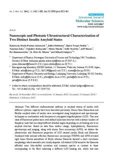
| dc.description.abstract | Two different conformational isoforms or amyloid strains of insulin with different cytotoxic capacity have been described previously. Herein these filamentous and fibrillar amyloid states of insulin were investigated using biophysical and spectroscopic techniques in combination with luminescent conjugated oligothiophenes (LCO). This new class of fluorescent probes has a well defined molecular structure with a distinct number of thiophene units that can adopt different dihedral angles depending on its binding site to an amyloid structure. Based on data from surface charge, hydrophobicity, fluorescence spectroscopy and imaging, along with atomic force microscopy (AFM), we deduce the ultrastructure and fluorescent properties of LCO stained insulin fibrils and filaments. Combined total internal reflection fluorescence microscopy (TIRFM) and AFM revealed rigid linear fibrous assemblies of fibrils whereas filaments showed a short curvilinear morphology which assemble into cloudy deposits. All studied LCOs bound to the filaments afforded more blue-shifted excitation and emission spectra in contrast to those corresponding to the fibril indicating a different LCO binding site, which was also supported by less efficient hydrophobic probe binding. Taken together, the multi-tool approach used here indicates the power of ultrastructure identification applying AFM together with LCO fluorescence interrogation, including TIRFM, to resolve structural differences between amyloid states. | nb_NO |
| dc.language.iso | eng | nb_NO |
| dc.publisher | MDPI | nb_NO |
| dc.relation.uri | http://www.mdpi.com/1422-0067/13/2/1461/pdf | |
| dc.title | Nanoscopic and Photonic Ultrastructural Characterization of Two Distinct Insulin Amyloid States | nb_NO |
| dc.type | Journal article | nb_NO |
| dc.type | Peer reviewed | en_GB |
| dc.date.updated | 2015-09-29T12:20:24Z | |
| dc.source.volume | 13 | nb_NO |
| dc.source.journal | International Journal of Molecular Sciences | nb_NO |
| dc.source.issue | 2 | nb_NO |
| dc.identifier.doi | 10.3390/ijms13021461 | |
| dc.identifier.cristin | 922922 | |
| dc.description.localcode | This is an open access article distributed under the Creative Commons Attribution License which permits unrestricted use, distribution, and reproduction in any medium, provided the original work is properly cited. | nb_NO |
