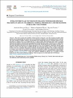
Intraventricular Vector Flow Imaging with Blood Speckle Tracking in Adults: Feasibility, Normal Physiology and Mechanisms in Healthy Volunteers
| dc.contributor.author | Daae, Annichen Søyland | |
| dc.contributor.author | Wigen, Morten Smedsrud | |
| dc.contributor.author | Fadnes, Solveig | |
| dc.contributor.author | Løvstakken, Lasse | |
| dc.contributor.author | Støylen, Asbjørn | |
| dc.date.accessioned | 2022-02-15T12:47:52Z | |
| dc.date.available | 2022-02-15T12:47:52Z | |
| dc.date.created | 2021-10-20T10:15:10Z | |
| dc.date.issued | 2021 | |
| dc.identifier.citation | Ultrasound in Medicine and Biology. 2021, 47 (12), 3501-3513. | en_US |
| dc.identifier.issn | 0301-5629 | |
| dc.identifier.uri | https://hdl.handle.net/11250/2979103 | |
| dc.description.abstract | This study examines the feasibility of blood speckle tracking for vector flow imaging in healthy adults and describes the physiologic flow pattern and vortex formation in relation to the wall motion in the left ventricle. The study included 21 healthy volunteers and quantified and visualized flow patterns with high temporal resolution down to a depth of 10–12 cm without the use of contrast agents. Intraventricular flow seems to originate during the isovolumetric relaxation with a propagation of blood from base to apex. With the E-wave, rapid inflow and vortex formation occurred on both sides of the valve basally. During diastasis the flow gathers in a large vortex before the pattern from the E-wave repeats during the A-wave. In isovolumetric contraction, the flow again gathers in a large vortex that seems to facilitate the flow out in the aorta during systole. No signs of a persistent systolic vortex were visualized. The geometry of the left ventricle and the movement of the AV-plane is important in creating vortices that are favorable for the blood flow and facilitate outflow. The quantitative measurements are in concordance with these findings, but the clinical interpretation must be evaluated in future clinical studies. | en_US |
| dc.language.iso | eng | en_US |
| dc.publisher | Elsevier | en_US |
| dc.rights | Navngivelse 4.0 Internasjonal | * |
| dc.rights.uri | http://creativecommons.org/licenses/by/4.0/deed.no | * |
| dc.title | Intraventricular Vector Flow Imaging with Blood Speckle Tracking in Adults: Feasibility, Normal Physiology and Mechanisms in Healthy Volunteers | en_US |
| dc.type | Peer reviewed | en_US |
| dc.type | Journal article | en_US |
| dc.description.version | publishedVersion | en_US |
| dc.source.pagenumber | 3501-3513 | en_US |
| dc.source.volume | 47 | en_US |
| dc.source.journal | Ultrasound in Medicine and Biology | en_US |
| dc.source.issue | 12 | en_US |
| dc.identifier.doi | 10.1016/j.ultrasmedbio.2021.08.021 | |
| dc.identifier.cristin | 1947204 | |
| cristin.ispublished | true | |
| cristin.fulltext | original | |
| cristin.qualitycode | 2 |
Files in this item
This item appears in the following Collection(s)

Except where otherwise noted, this item's license is described as Navngivelse 4.0 Internasjonal