| dc.contributor.author | Hansen, Tor Ivar | |
| dc.contributor.author | Brezova, Veronika | |
| dc.contributor.author | Eikenes, Live | |
| dc.contributor.author | Håberg, Asta | |
| dc.contributor.author | Vangberg, Torgil Riise | |
| dc.date.accessioned | 2021-09-24T10:44:33Z | |
| dc.date.available | 2021-09-24T10:44:33Z | |
| dc.date.created | 2015-10-05T14:05:09Z | |
| dc.date.issued | 2015 | |
| dc.identifier.citation | American Journal of Neuroradiology. 2015, 36 (8), 1450-1456. | en_US |
| dc.identifier.issn | 0195-6108 | |
| dc.identifier.uri | https://hdl.handle.net/11250/2781375 | |
| dc.description.abstract | BACKGROUND AND PURPOSE: The intracranial volume is commonly used for correcting regional brain volume measurements for variations in head size. Accurate intracranial volume measurements are important because errors will be propagated to the corrected regional brain volume measurements, possibly leading to biased data or decreased power. Our aims were to describe a fully automatic SPM-based method for estimating the intracranial volume and to explore the practical implications of different methods for obtaining the intracranial volume and normalization methods on statistical power.
MATERIALS AND METHODS: We describe a method for calculating the intracranial volume that can use either T1-weighted or both T1- and T2-weighted MR images. The accuracy of the method was compared with manual measurements and automatic estimates by FreeSurfer and SPM-based methods. Sample size calculations on intracranial volume–corrected regional brain volumes with intracranial volume estimates from FreeSurfer, SPM, and our proposed method were used to explore the benefits of accurate intracranial volume estimates.
RESULTS: The proposed method for estimating the intracranial volume compared favorably with the other methods evaluated here, with mean and absolute differences in manual measurements of −0.1% and 2.2%, respectively, and an intraclass correlation coefficient of 0.97 when using T1-weighted images. Using both T1- and T2-weighted images for estimating the intracranial volume slightly improved the accuracy. Sample size calculations showed that both the accuracy of intracranial volume estimates and the method for correcting the regional volume measurements affected the sample size.
CONCLUSIONS: Accurate intracranial volume estimates are most important for ratio-corrected regional brain volumes, for which our proposed method can provide increased power in intracranial volume–corrected regional brain volume data. | en_US |
| dc.language.iso | eng | en_US |
| dc.publisher | American Society of Neuroradiology | en_US |
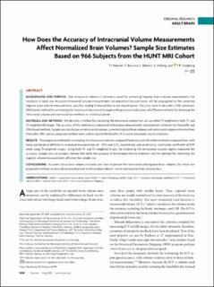
| dc.title | How Does the Accuracy of Intracranial Volume Measurements Affect Normalized Brain Volumes? Sample Size Estimates Based on 966 Subjects from the HUNT MRI Cohort | en_US |
| dc.type | Peer reviewed | en_US |
| dc.type | Journal article | en_US |
| dc.description.version | publishedVersion | en_US |
| dc.rights.holder | © 2015 by American Journal of Neuroradiology Indicates open access to non-subscribers at www.ajnr.org | en_US |
| dc.source.pagenumber | 1450-1456 | en_US |
| dc.source.volume | 36 | en_US |
| dc.source.journal | American Journal of Neuroradiology | en_US |
| dc.source.issue | 8 | en_US |
| dc.identifier.doi | 10.3174/ajnr.A4299 | |
| dc.identifier.cristin | 1278465 | |
| cristin.ispublished | true | |
| cristin.fulltext | original | |
| cristin.qualitycode | 1 | |